

 Open Access||Case Report
Fatal Necrotizing and Suppurative Masticatory Myositis with Intracranial Involvement in an Albino Bennett's Wallaby (Notamacropus rufogriseus)
Andreia Garcês1,2,3 , Luís Sousa1,3, Camila Cardoso1,3, Roberto Sargo1,2,3, Luana Monteiro1, Diogo Silva1, Ricardo Lopes3,4,5, Filipe Silva1,2,3, Justina Prada2,3
1 Wildlife Rehabilitation Centre (CRAS), Veterinary Teaching Hospital, University of Trás-os-Montes e Alto Douro (UTAD), Quinta dos Prados, 4500-801, Vila Real, Portugal
2 Associate Laboratory for Animal and Veterinary Sciences (AL4AnimalS), Animal and Veterinary Research Centre (CECAV), University of Trás-os-Montes e Alto Douro (UTAD), Quinta dos Prados, 4500-801, Vila Real, Portugal
3 Department of Veterinary Sciences, School of Agrarian and Veterinary Sciences (ECAV), University of Trás-os-Montes e Alto Douro (UTAD), Quinta dos Prados, 4500-801, Vila Real, Portugal
4 Department of Veterinary and Animal Sciences, University Institute of Health Sciences (IUCS), CESPU, 4585-116 Gandra, Portugal
5 CEDIVET Veterinary Laboratories, Lionesa Business Hub, R. Lionesa 446 C24, 4465-671 Leça do Balio, Portugal
Abstract
A juvenile female Bennett's wallaby (Notamacropus rufogriseus) was presented in an unconscious state and died shortly after admission. Post-mortem examination revealed severe necrotizing and suppurative myositis affecting the masticatory muscles, with extensive abscess formation and associated subcutaneous hematoma. Purulent material extended from the affected soft tissues toward the optic canal and cranial cavity, and a subdural hematoma was also identified. Histopathological examination confirmed extensive myofiber degeneration and necrosis, marked suppurative inflammation, tissue disruption, and numerous intralesional bacterial colonies. Microbiological culture of the affected tissues yielded Enterobacter cloacae complex and Enterococcus faecalis, supporting a polymicrobial bacterial infection. Based on the distribution and characteristics of the lesions, the infection was considered most likely to have originated from trauma to the oral soft tissues, followed by local bacterial invasion and progressive extension into adjacent structures. Unlike the more commonly reported chronic "lumpy jaw" syndrome of macropods, this case represents an acute and rapidly progressive bacterial soft-tissue infection involving the masticatory musculature. The findings emphasize the value of comprehensive post-mortem examination, histopathology, microbiological culture, and antimicrobial susceptibility testing for accurately characterizing jaw-associated disease in macropods.
Keywords
Graphical Abstract
Novelty Statement
This study reports a novel, rapidly progressive phenotype of macropod jaw disease: an acute, fatal polymicrobial masticatory myositis with cranial extension in a Bennett's wallaby (Notamacropus rufogriseus) that challenges the traditional chronic 'lumpy jaw' paradigm.
1. Introduction
Bennett's wallaby (Notamacropus rufogriseus) is a medium-sized marsupial native to the temperate forests and grasslands of eastern Australia and Tasmania.[1] Recognized by its reddish-brown fur and lighter underparts, this species is mainly nocturnal and herbivorous, feeding on grasses and leaves.[1] Bennett's wallabies are known for their adaptability to various habitats and climates. It is a species common in zoological collections, due to its manageable size, gentle nature, and ease of breeding in captivity. It is considered to have a status of Least Concern according to the IUCN Red List of Threatened Species.[2]
There are a few documented pathologies involving the jaw/masticatory region in wallabies. Common issues reported include myositis, muscle abscesses, fibrosis, and traumatic injury resulting from fighting, capture, or feeding behavior. Nutritional myopathy (white muscle disease), caused by selenium or vitamin E deficiency, and "lumpy jaw" caused by bacterial infections, such as Actinomyces spp. or Fusobacterium spp., have been reported in this species.[3,4]
To the authors' knowledge, this case represents an uncommon presentation of acute necrotizing and suppurative masticatory myositis in a Bennett's wallaby (N. rufogriseus, Desmarest, 1817), with bacterial isolation and suspected intracranial extension, in the absence of gross mandibular or maxillary osteolysis.
2. Case presentation
A juvenile female Bennett's wallaby (N. rufogriseus) was admitted to the Veterinary Hospital of UTAD in a hypothermic and unconscious state, exhibiting haemoptysis and convulsive activity. This case report was prepared in accordance with the CARE guidelines, where applicable. Given the presence of haemoptysis and the initially undetermined aetiology, standard veterinary biosecurity precautions, including appropriate personal protective equipment and routine contamination-control procedures, were applied during clinical handling and post-mortem examination. Instruments, surfaces, biological waste, and the carcass were subsequently managed in accordance with the Veterinary Hospital institutional biosecurity and disposal procedures.
Although the animal was in critical condition, emergency stabilization was considered appropriate because a potentially treatable condition could not be excluded at the initial assessment. Intravenous access was therefore immediately attempted to enable supportive and symptomatic treatment. However, the animal suffered cardiopulmonary arrest and died during placement of the intravenous catheter, only a few minutes after admission, before further diagnostic evaluation or pharmacological treatment, prognostic reassessment, or consideration of euthanasia could be undertaken.
Following death, the carcass was refrigerated at approximately 5°C and was not frozen prior to post-mortem examination the following day. The necropsy was conducted in accordance with standard veterinary pathology procedures and institutional practices. A systematic external and internal examination was performed, with appropriate photographic documentation and collection of representative tissue samples for histopathological and microbiological analyses.
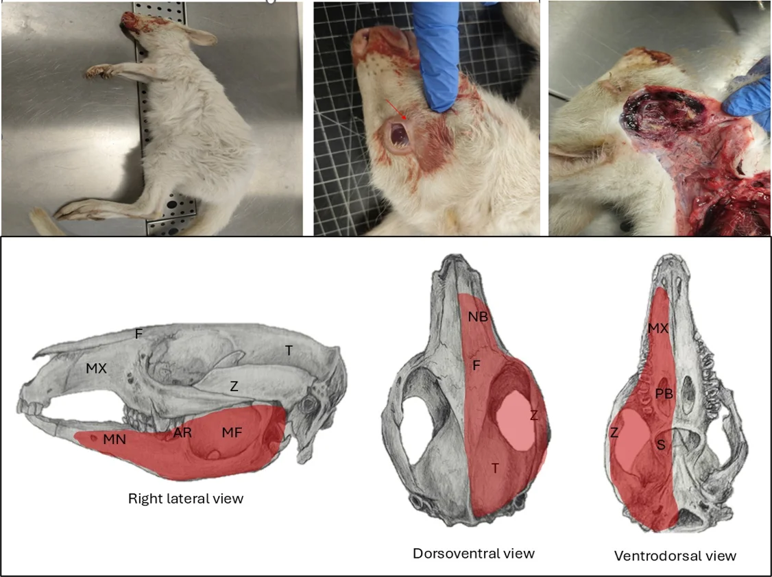
On external examination, the carcass was in good body condition (score 3/5). Fresh, unclotted blood was present around the oral cavity and adhered to the surrounding fur. The right periocular region exhibited mild oedema, and a small amount of serous discharge was noted from the palpebral fissure (Fig. 1).
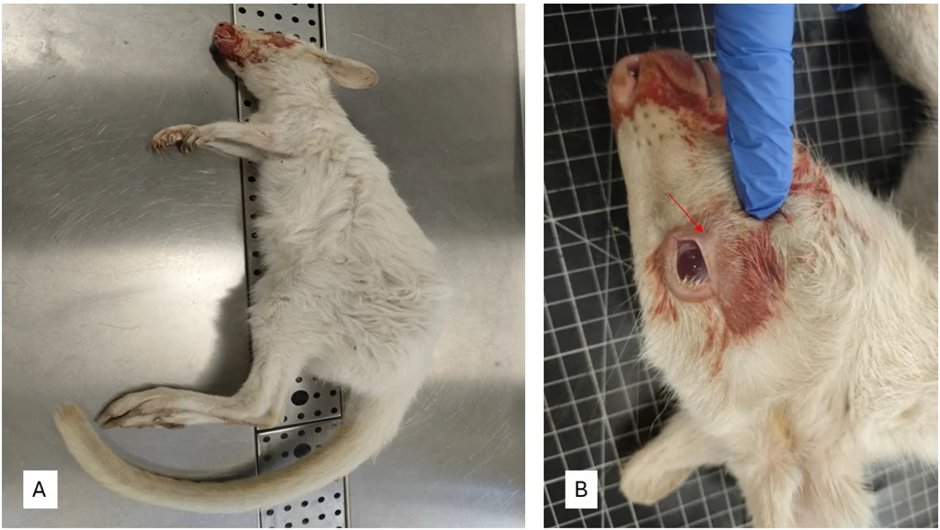
Fig. 1: (A) Juvenile albino female Bennett's wallaby (N. rufogriseus); (B) Right periocular region showing mild edema and serous discharge (red arrow).
The oral cavity emitted a fetid odour upon opening. Following reflection of the skin, a well-defined subcutaneous and intramuscular hematoma was observed over the right thoracic region, involving the pectoralis musculature and overlying the ribs. The right masseter and digastric muscles exhibited moderate oedema and marked vascular congestion (Fig. 2A). On cut section, the muscles were diffusely dark red to black, friable, with extensive areas of necrosis. The cut surface exuded bloody fluid admixed with small pockets of yellow purulent material (Fig. 2B), consistent with severe necrotizing and haemorrhagic myositis. The orbicularis oris muscle showed dark red discoloration, friable to the touch, and easily detached from the underlying bone, indicating acute myonecrosis (Fig. 2C).
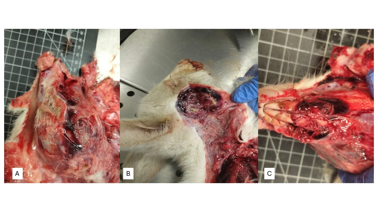
Fig. 2: (A) Gross lesions of the right masticatory region. Moderate edema and marked vascular congestion of the right masseter and digastric muscles. (B) Dark red to black, friable muscle with extensive necrosis and purulent foci. (C) Dark red discoloration and friability of the orbicularis oris muscle.
Adjacent to the masseteric fossa, a focal lesion approximately 7×4 cm in size was identified, exhibiting a yellow discolouration and a pasty consistency, findings consistent with a localized purulent inflammatory process. Purulent exudate was observed extending through the adjacent soft tissues into the optic canal and toward the cranial cavity. The adjacent bone appeared intact, with no evidence of osteolysis or fracture (Fig. 3).
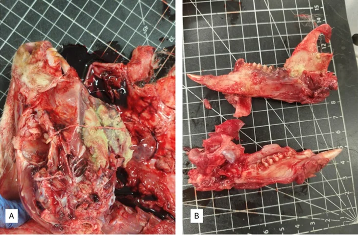
Fig. 3: Abscess formation near the masseteric fossa. (A) Purulent lesion adjacent to the masseteric fossa. (B) Abscess overlying the jawbone; no gross bone destruction was observed. The contralateral healthy jaw is shown for comparison.
The cerebrum displayed a dark discoloration of the right hemisphere, and upon opening of the meninges, a hematoma was evident within the subdural space. The meningeal vessels were markedly congested, and the cerebral parenchyma had a softened consistency and was diffusely congested (Fig. 4).
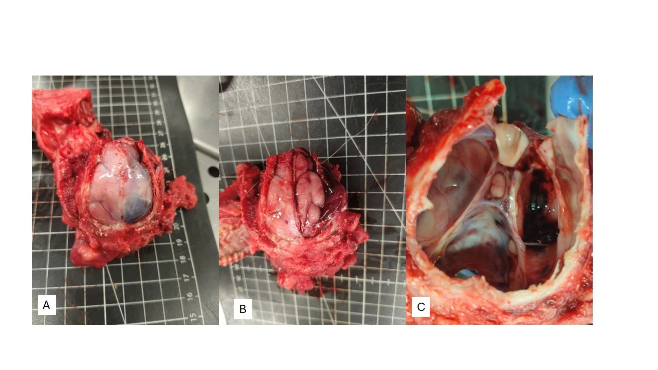
Fig. 4: Intracranial and perioptic lesions. (A) Hematoma affecting the right cerebral hemisphere. (B) Subdural hematoma. (C) Hemorrhagic and suppurative material extending toward the optic canal.
Fig. 5 is a schematic representation of the affected areas.
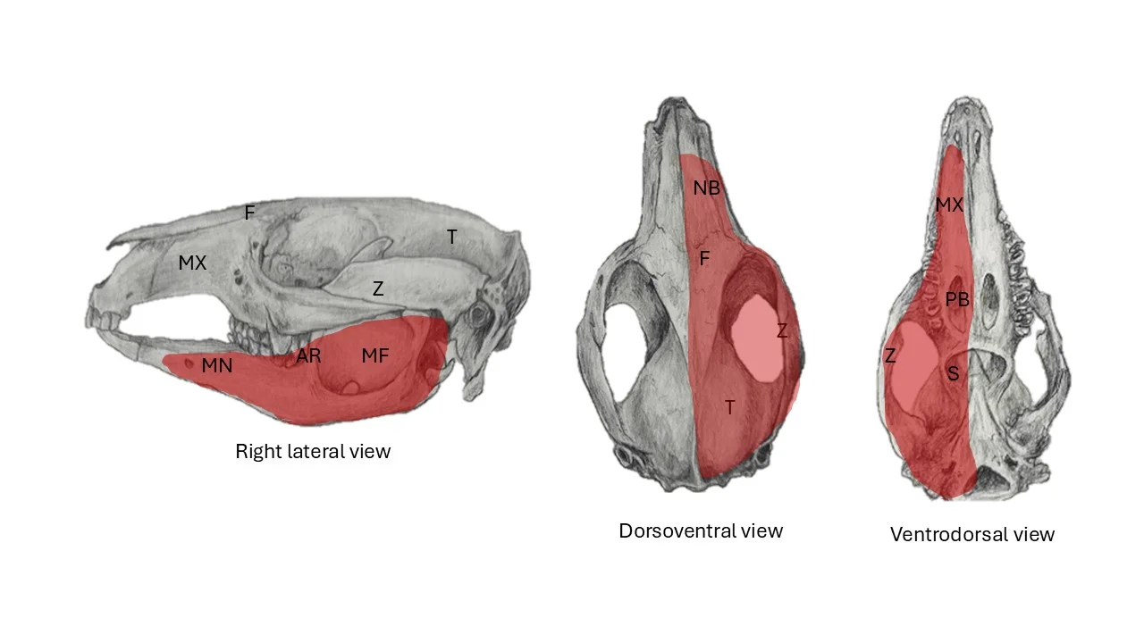
Fig. 5: Schematic representation of the affected anatomical regions, marked in red. MX: maxilla; MN: mandible; AR: anterior border of the ascending ramus of the dentary; MF: masseteric fossa; Z: zygomatic bone; F: frontal bone; NB: nasal bone; T: temporal bone; PB: palatine bone; S: sphenoid bone. Source: Illustration by Andreia Garcês.
Following completion of the post-mortem examination and diagnostic sampling, the carcass was incinerated in accordance with the standard procedures of the Veterinary Hospital. Samples of the masticatory muscle, brain, meninges, and optic canal were submitted for histopathology examination. The tissues were fixed in 10% neutral-buffered formalin and stained with H&E. Microscopic examination of the affected masticatory muscles (masseter and digastric) revealed extensive necrosis of myofibers with loss of cross-striations and fragmentation. There was a dense mixed inflammatory infiltrate composed of neutrophils, macrophages, lymphocytes, and plasma cells. Multifocally, intralesional bacterial colonies composed of coccoid and bacillary forms were observed. The surrounding connective tissue and viable muscle fibers exhibited oedema, vascular congestion, and focal thrombosis, and mild mononuclear infiltration.
Samples of purulent tissue were cultured on Blood Agar, Columbia Nalidixic Acid (CNA) Agar, and MacConkey Agar (BioMérieux, France) and incubated at 35–37°C for 48 hours. Aerobic and anaerobic bacterial cultures were performed. Aerobic cultures were incubated at 35–37°C for 48 h, whereas anaerobic cultures were incubated at 37°C under anaerobic conditions using an anaerobic jar with a GasPak for 72 h. Bacterial identification was performed using the VITEK® system (2 GP ID Card REF 21342; 2 GN ID Card REF 21341, bioMérieux, France). Aerobic culture yielded Enterobacter cloacae complex and Enterococcus faecalis. No strict anaerobic bacteria were isolated. Antimicrobial susceptibility was interpreted according to EUCAST criteria.
The Enterobacter cloacae complex isolate was susceptible to amikacin, cefpodoxime, ceftiofur, doxycycline, enrofloxacin, gentamicin, imipenem, marbofloxacin, neomycin, pradofloxacin, tetracycline, and trimethoprim/sulfamethoxazole. It showed resistance to amoxicillin/clavulanic acid, ampicillin, cephalexin, and cephalothin, and intermediate susceptibility to cefovecin and chloramphenicol.
The Enterococcus faecalis isolate was susceptible to benzylpenicillin, chloramphenicol, enrofloxacin, and marbofloxacin. It was resistant to doxycycline and tetracycline and showed intermediate susceptibility to erythromycin. Antimicrobial susceptibility testing was performed and interpreted according to the European Committee on Antimicrobial Susceptibility Testing (EUCAST) Clinical Breakpoint Tables, version v14.0 (2024).
3. Results and discussion
The juvenile Bennett's wallaby presented with severe necrotizing and suppurative myositis of the masticatory muscles, associated with abscess formation, subcutaneous hematoma, and intracranial involvement. Histopathology and bacterial culture supported a diagnosis of acute polymicrobial bacterial myositis involving Enterobacter cloacae complex and Enterococcus faecalis.[5]
The most likely portal of entry may be oral soft-tissue trauma, possibly caused by coarse vegetation or hard food material. Such trauma may have allowed opportunistic bacteria to colonize damaged tissues and spread through adjacent fascial planes. Similar jaw-associated infections have been reported in macropods and are often associated with oral trauma, opportunistic bacterial colonization, and secondary osteomyelitis or myositis.[5-7]
Although some features overlapped with classic lumpy jaw, the present case showed important differences. Lumpy jaw in macropods is usually a chronic granulomatous osteomyelitis of the mandible or maxilla, frequently associated with periosteal proliferation, bone lysis, sulphur granules, and bacteria such as Actinomyces spp. In contrast, this case was characterized by acute necrotizing and suppurative soft-tissue infection, rapid clinical deterioration, and no gross evidence of osteolysis or fracture. These findings support acute bacterial myositis as the primary pathological process rather than typical chronic lumpy jaw.[4,8]
The anatomical continuity between the purulent lesion, optic canal, and cranial cavity suggests that infectious extension may have contributed to the intracranial findings. However, the subdural hematoma should be interpreted cautiously, as trauma during collapse, convulsions, or handling may also have played a role.[4,8]
4. Conclusion
This case highlights the importance of complete post-mortem examination, histopathology, microbiological culture, and antimicrobial susceptibility testing in macropod jaw disease. Acute necrotizing soft-tissue infections should be considered as differential diagnoses for jaw-associated lesions in wallabies, particularly when rapid clinical deterioration occurs and gross bone involvement is absent.
Acknowledgement
Not applicable.
CRediT Author Contribution Statement
Andreia Garcês: Conceptualization, Methodology, Formal Analysis, Investigation, Validation, Visualization, Supervision, Project Administration, Writing – Original Draft, Writing – Review & Editing. Luís Sousa: Data curation, Resources, Investigation, Software. Camila Cardoso: Data curation, Resources, Investigation, Software. Roberto Sargo: Data curation, Resources, Investigation, Software. Luana Monteiro: Data curation, Resources, Investigation, Software. Diogo Silva: Data curation, Resources, Investigation, Software. Ricardo Lopes: Data curation, Resources, Investigation, Software. Filipe Silva: Supervision, Writing – Review & Editing. Justina Prada: Conceptualization, Methodology, Formal Analysis, Investigation, Validation, Visualization, Supervision, Project Administration, Writing – Original Draft, Writing – Review & Editing. All authors have read and approved the final version of the manuscript for publication and agree to be accountable for all aspects of the work, ensuring that questions related to the accuracy or integrity of any part of the work are appropriately investigated and resolved.
Funding
This work was supported by the Portuguese Foundation for Science and Technology (Fundação para a Ciência e a Tecnologia, FCT) under grant numbers UIDB/CVT/00772/2020 and LA/P/0059/2020, awarded to CECAV. The funder had no role in the study design, data collection, data analysis or interpretation, preparation of the manuscript, or decision to submit the work for publication.
Institutional Review Board Statement
This study describes a retrospective clinical case involving routine emergency veterinary care and diagnostic post-mortem examination, without experimental intervention. Formal institutional ethical approval was therefore not required. All procedures were conducted in accordance with the institutional standards of the Veterinary Hospital of the University of Trás-os-Montes and Alto Douro (UTAD) and applicable national animal welfare legislation.
Informed Consent Statement
The Bennett's wallaby was a privately owned animal and was legally maintained in accordance with applicable national requirements. Informed consent was obtained from the animal's legal owner/custodian for emergency clinical care and subsequent post-mortem examination.
Consent to Publish Statement
Explicit consent was also obtained for publication of the clinical data, pathological findings, and associated images presented in this case report. All identifying information relating to the animal's owner or legal custodian, as well as precise geographic details that could permit identification of the animal's origin or location, was omitted to preserve client confidentiality.
Data Availability Statement
The datasets generated and/or analyzed during the current study that support the findings are available from the corresponding author upon reasonable request.
Conflict of Interest
There is no conflict of interest.
Artificial Intelligence (AI) Use Disclosure
The authors declare that artificial intelligence (AI)-assisted tools were used only for language refinement, grammar improvement, and manuscript structuring purposes during the preparation of this work. All technical content, experimental implementation, results, and interpretations were independently developed and verified by the authors.
Supporting Information
Not applicable.
References
- [1] W. Clemens, B. Richardson, P. Baverstock, Fauna of Australia, Fauna of Australia series, ABRS Publications, 1989, 1B.
- [2] N. McKenzie, P. Menkhorst, D. Lunney, Macropus rufogriseus. The IUCN Red List of Threatened Species 2016: e.T40566A21953329, 2016, doi: 10.2305/IUCN.UK.2016-2.RLTS.T40566A21953329.en, Accessed 29 July 2026.
- [3] I. S. Braga, S. Tanaka, K. Ochiai, C. Itakura, Suspected nutritional myopathy in two captive Bennett's wallabies (Macropus rufogriseus), Japanese Journal of Veterinary Research, 1995, 43, 99–103, doi: 10.14943/jjvr.43.2.99.
- [4] J. L. Samuel, Jaw disease in macropod marsupials: bacterial flora isolated from lesions and from the mouths of affected animals, Veterinary Microbiology, 1983, 8, 373–387, doi: 10.1016/0378-1135(83)90050-0.
- [5] M. D. Brookins, S. Rajeev, T. D. Thornhill, K. Kreinheder, D. L. Miller, Mandibular and maxillary osteomyelitis and myositis in a captive herd of red kangaroos (Macropus rufus), Journal of Veterinary Diagnostic Investigation, 2008, 20, 846–849, doi: 10.1177/104063870802000627.
- [6] L. P. Kane, J. N. Langan, M. J. Adkesson, S. K. Chinnadurai, B. N. Nevitt, R. Drees, Treatment of mandibular osteomyelitis in two red-necked wallabies (Macropus rufogriseus) by means of intensive long-term parenteral drug administration and serial computed tomographic monitoring, Journal of the American Veterinary Medical Association, 2017, 251, 1070–1077, doi: 10.2460/javma.251.9.1070.
- [7] N. Hoyer, J. Rawlinson, E. Klaphake, Treatment of oral disease in eight captive Bennett's wallabies (Macropus rufogriseus) between 2011 and 2019: a case series, Journal of Zoo and Wildlife Medicine, 2020, 51, 705–719, doi: 10.1638/2019-0139.
- [8] M. R. H. Taylor, P. Wilson, L. Smyth, L. English, C. T. Keane, Laboratory investigations in the "lumpy jaw" of macropods, Veterinary Science Communications, 1978, 2, 131–136, doi: 10.1007/BF02291441.