

 Open Access||Research Article
Development of a Latex Agglutination Test (LAT) using Whole-Cell Protein for the Detection of Mycoplasma ovipneumoniae Antibodies in Small Ruminants
Shanmugasundaram Udhayavel1 , Kuppannan Sukumar2, Kuppusamy Senthilkumar3, Palani Srinivasan4, Ayyasamy Elango5
1 Department of Veterinary Microbiology, Veterinary College and Research Institute, Tamil Nadu Veterinary and Animal Sciences University, Salem, Tamil Nadu, 636112, India
2 Department of Veterinary Microbiology, Veterinary College and Research Institute, Tamil Nadu Veterinary and Animal Sciences University, Namakkal, Tamil Nadu, 637002, India
3 Centralised Clinical Laboratory, Madras Veterinary College, Veterinary and Animal Sciences University, Chennai, Tamil Nadu, 600007, India
4 Poultry Disease Diagnosis and Surveillance Laboratory, Veterinary College and Research Institute campus, Tamil Nadu Veterinary and Animal Sciences University, Namakkal, Tamil Nadu, 637002, India
5 Veterinary College and Research Institute, Tamil Nadu Veterinary and Animal Sciences University, Salem, Tamil Nadu, 636112, India
Abstract
Mycoplasma ovipneumoniae is a major pathogen responsible for respiratory illness in sheep and goats, particularly chronic nonprogressive pneumonia, and its presence results in substantial economic losses within the small ruminant sector. Although diagnostic methods such as culture and polymerase chain reaction (PCR) are routinely used, each approach has inherent limitations. There is no rapid, inexpensive and field-deployable serological assay available for the detection of M. ovipneumoniae antibodies in small ruminants. The objective of the present study was to develop a latex agglutination test (LAT) to detect Mycoplasma ovipneumoniae antibodies in small ruminants at the field level. A latex agglutination test (LAT) was developed using whole-cell protein antigen extracted from M. ovipneumoniae coupled with latex beads (0.80 µm) and tested on serum samples from 33 PCR-positive and 77 PCR-negative animals. A comparison of the latex agglutination test results with those of PCR revealed 93.94% sensitivity and 93.51% specificity. The developed serological assay strongly agreed with the PCR results, with a kappa value greater than 0.81. Hence, this latex agglutination test kit offers a convenient and rapid means of diagnosing infections in field settings and is particularly useful for large-scale surveillance of infections in flocks.
Keywords
Graphical Abstract
Novelty Statement
This study introduces the first rapid, low-cost, and penside Latex Agglutination Test (LAT) for detecting Mycoplasma ovipneumoniae antibodies directly in small ruminant field settings. By utilizing a whole-cell protein antigen, this novel assay achieves over 93% sensitivity and specificity, offering a highly accessible tool for large-scale flock surveillance without requiring complex laboratory infrastructure.
1. Introduction
Sheep and goats play a vital role in the livelihoods of Indian farmers by contributing substantially to meat and milk production and by exhibiting rapid growth rates. Consequently, they are considered valuable economic assets. Respiratory diseases in these animals lead to reduced weight gain, increased mortality and considerable financial losses.[1] Such infections arise from multiple interacting factors, with a wide range of etiological agents contributing to the respiratory disease complex.[2,3] Among these, mycoplasmosis, an emerging transboundary disease of sheep and goats, imposes heavy economic burdens on farmers and disrupts trade in many regions.[4-6] Mycoplasma ovipneumoniae is among the most important pathogens associated with chronic, nonprogressive pneumonia in small ruminants. First reported in 1963 in Scotland in sheep with pulmonary adenomatosis, the organism has since been identified in both clinically affected and apparently healthy animals.[7] Molecular detection of the organism has been reported in various parts of India: isolates from pneumonic sheep and goats in Andhra Pradesh have been confirmed using PCR on nasal swabs, while Santhiya et al., identified the pathogen in goats exhibiting respiratory symptoms in northern and central Kerala.[8,9] Similarly, M. ovipneumoniae has been detected in nasal secretions, tissues and synovial fluids of symptomatic small ruminants in the Bengaluru region of Karnataka.[10] These reports emphasize the need for accurate and prompt diagnosis to implement appropriate control measures.
Although diagnostic methods such as culture, polymerase chain reaction (PCR) and enzyme-linked immunosorbent assay (ELISA) are widely utilized, each has inherent drawbacks. The culture is labor intensive, requires stringent growth conditions, and often fails because of the fastidious nature of Mycoplasma species. PCR provides superior sensitivity and specificity but is costly, depends on advanced laboratory facilities, and is impractical for large-scale screening in field settings. ELISA is more user friendly, yet its reliability may fluctuate because antigens can cross-react with related Mycoplasma spp. The latex agglutination test (LAT) has been explored historically as a rapid serodiagnostic tool. Kende reported that LAT performed comparably to tetrazolium reduction inhibition and complement fixation tests (CFT) for detecting M. pneumoniae antibodies.[11] Slavik and Switzer applied LAT to diagnose M. hyopneumoniae infection in pigs and observed persistent LAT antibody responses for up to 48 weeks.[12] Rurangirwa et al., developed a polysaccharide-based LAT for diagnosing contagious caprine pleuropneumonia (CCPP), which has a higher sensitivity than CFT does and is suitable as a pen-side test with results available in minutes.[13] Subsequent applications in various regions have demonstrated the practicality and cost-effectiveness of LAT for screening for mycoplasmal infections.[14-17] Despite these advancements, no rapid, low-cost, field-ready serological assay currently exists for detecting antibodies to M. ovipneumoniae in sheep and goats. LAT represents a promising alternative because of its simplicity, affordability and minimal equipment requirements. However, a dedicated LAT kit for M. ovipneumoniae has yet to be developed. Establishing such a test would enable quick pen-side screening, enhance surveillance programs and support timely management of respiratory diseases in small ruminant populations.
2. Materials and methods
2.1 Development and standardization of the Latex Agglutination Test kit
The latex bead-sensitized whole-cell protein of Mycoplasma ovipneumoniae was prepared by the method outlined by Sankar et al., with slight modifications.[16]
2.2 Preparation of whole-cell protein
A reference isolate of Mycoplasma ovipneumoniae, which was isolated in our earlier study (Udhayavel et al.,) was initially inoculated into 2 ml of Mycoplasma experience liquid medium and incubated at 37°C for three days.[18] The inoculum was then transferred to 100 ml of medium and incubated at 37°C for an additional two days. Subculturing was subsequently performed in 500 ml of Mycoplasma Experience liquid medium, which was subsequently incubated at 37°C for three days with intermittent shaking. Afterward, the Mycoplasma cells were harvested by centrifugation at 18,000 rpm in a cooling centrifuge for one hour. Sterility checks were regularly conducted with each passage to prevent bacterial contamination. The cells were subsequently washed three times in sterile PBS, resuspended in PBS, and then sonicated with constant pulses for 30 seconds, with 30-second breaks, which was repeated 19 times. The concentration of whole-cell protein was estimated using the Bradford method.
2.3 Sensitization of latex beads with whole-cell protein
Latex beads (Sigma, 0.80 µm) were used. The latex bead suspension (10%) was washed twice by centrifugation at 6700 × g for three minutes each time in carbonate–bicarbonate buffer. The beads were then made into a 2% suspension with carbonate-bicarbonate buffer that was later mixed with an equal volume of M. ovipneumoniae whole-cell protein antigen (20 µg/ml) diluted in the same buffer. This mixture was incubated at 37°C for six hours with constant shaking at 250 × g. The sensitized beads were subsequently centrifuged at 6700 × g for three minutes, after which the pellet was resuspended as a 2% suspension in PBS containing 5 mg/ml bovine serum albumin (BSA; Himedia). The latex beads were then left at 37°C in a water bath overnight. Finally, the beads were centrifuged again as before, and the pellet was resuspended in PBS containing 0.5 mg/ml BSA.
2.4 Latex Agglutination Test kit
A volume of 25 µl of suspected serum was placed onto a clean glass slide. Similarly, 25 µl of the prepared sensitized latex beads coated with Mycoplasma ovipneumoniae whole-cell antigen was placed adjacent to the serum. The serum samples collected from the animals whose PCR results were positive and negative in our earlier study (Udhayavel et al.,) and the antigen were mixed using a stirring rod to form a circular area approximately 1.5 cm in diameter.[18] The glass plate was gently rocked for three minutes.
2.5 Assessment of the diagnostic characteristics of the developed Latex Agglutination Test kit
A latex agglutination test was performed on serum samples collected from 33 PCR-positive and 77 PCR-negative animals. The results of the latex agglutination test were compared with those of the PCR assay to determine indices such as sensitivity, specificity and Cohen's kappa coefficient (k) statistics, with PCR used as the gold standard. MedCalc's diagnostic test evaluation calculator was used. The strength of agreement determined by Cohen's kappa coefficient (k) was interpreted using standard benchmarks: k ≤ 0 indicates no agreement, 0.01–0.20 slight, 0.21–0.40 fair, 0.41–0.60 moderate, 0.61–0.80 substantial, and 0.81–1.00 indicates almost perfect agreement.
3. Results
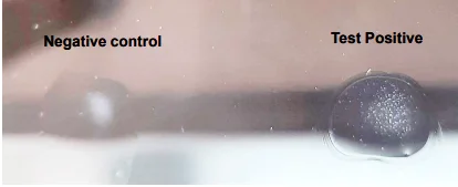
A latex agglutination test was performed on serum samples collected from 33 Mycoplasma ovipneumoniae-specific PCR-positive and 77 PCR-negative animals. Among the 110 corresponding serum samples tested, 31 showed agglutination when the latex agglutination kit was used (Fig. 1). Two nasal swab samples tested positive by PCR, but the corresponding serum samples did not show agglutination in the LAT region. Similarly, five nasal swabs were negative according to PCR, but when the corresponding serum samples were tested, they were positive according to the latex agglutination test. Therefore, among the 110 serum samples screened, 36 (32.73%) tested positive by the latex agglutination test. The results are shown in Table 1.
Fig. 1: Latex agglutination test for the detection of Mycoplasma ovipneumoniae antibodies. Note: Agglutination with Latex beads sensitized with Mycoplasma ovipneumoniae whole-cell protein.
Table 1: Comparative evaluation of the latex agglutination test with PCR
| PCR Positive | PCR Negative | Total | ||
|---|---|---|---|---|
| Latex agglutination test | Positive | 31 (a) | 5 (b) | 36 (a+b) |
| Negative | 2 (c) | 72 (d) | 74 (c+d) | |
| Total | 33 (a+c) | 77 (b+d) | 110 |
Note: a - true positive, b - false positive, c - false negative, d - true negative
3.1 Assessment of the diagnostic characteristics of the developed Latex Agglutination Test kit
The sensitivity and specificity of the latex agglutination test compared with those of PCR were 93.94 and 93.51%, respectively. Kappa statistics, which were used to analyze the agreement between the two tests, revealed a score of 0.852, indicating a high level of agreement (Table 2).
Table 2: Diagnostic characteristics of the latex agglutination test
| Diagnostic characteristics | Formula | Value |
|---|---|---|
| Sensitivity | [a/(a + c)] × 100 | 93.94% (95% CI: 79.82%–99.34%) |
| Specificity | [d/(b + d)] × 100 | 93.51% (95% CI: 85.50%–97.85%) |
| Kappa value | k= a+d-P/1-P | 0.852 ± 0.054 (SE) |
| Statistical Significance (p-value)* | Fisher's Exact Test | p < 0.0001 |
Note: An association is considered highly statistically significant when p < 0.05.
4. Discussion
The plate agglutination test primarily detects animals in the early (acute) stage of the disease.[19] The plate agglutination test requires larger bacterial cells to form visible aggregates. By attaching Mycoplasma cells to carrier particles such as latex, the mass of the cells can be increased, and the specificity of the Mycoplasma surface antigens can be maintained.[20] The latex agglutination test is a simple macroagglutination test that combines sensitivity with affordability and ease of use under field conditions, eliminating the need for specialized training or equipment. In this study, a latex agglutination test (LAT) was performed using latex beads (0.8 µm diameter) coated with sonicated Mycoplasma ovipneumoniae whole-cell protein. Ramadass et al., employed latex beads with a diameter of 0.8 µm for an agglutination test and reported that the test results were influenced by the size of the latex particles.[21] In this study, carbonate–bicarbonate buffer was used to dilute the latex particles during the sensitization of the beads. This approach aligns with the findings of Ramadass et al., who also identified this buffer as suitable for latex bead sensitization.[14] Sankar et al., also developed LAT based on whole-cell protein antigens to detect Mycoplasma gallisepticum antibodies from chicken serum in Kerala.[16] In the current study, the results of LAT were compared with those of PCR. Among the 110 serum samples tested, 31 showed agglutination in the LAT region. Two false negative and five false positive results were obtained. Hence, the seropositivity of M. ovipneumoniae antibodies in the latex agglutination test was 32.73% (36/110). The sensitivity and specificity of the newly developed LAT kit were determined to be 93.94% and 93.51%, respectively. Furthermore, the calculated Cohen's kappa coefficient was 0.852, which falls within the 0.81–1.00 range. This statistically demonstrates an almost perfect agreement with the reference PCR gold standard, confirming the diagnostic robustness and reliability of the assay. This finding is in accordance with the findings of Sankar et al. (2013), who reported a sensitivity and specificity of 95.24% and 93.33%, respectively.[16] Hence, this agglutination-based kit offers a convenient and rapid means of diagnosing infections in field settings and is particularly useful for large-scale surveillance of infections in flocks rather than individual testing.
5. Conclusion
The latex agglutination test (LAT) developed in the present study demonstrated good diagnostic performance for the detection of Mycoplasma ovipneumoniae antibodies in small ruminants. The test was simple, rapid and easy to perform, with minimal technical requirements, making it suitable for routine screening of sheep and goat flocks. The satisfactory sensitivity and specificity observed in the study indicate that the LAT can serve as a useful preliminary serological screening tool for identifying animals exposed to M. ovipneumoniae. Its simplicity and rapid visual interpretation also offer advantages for application under field conditions, particularly in areas where sophisticated laboratory facilities are not readily available.
Acknowledgement
The authors thank the Tamil Nadu Veterinary and Animal Sciences University, Chennai, India.
CRediT Author Contribution Statement
Shanmugasundaram Udhayavel: Investigation, Data Curation, Formal Analysis, Writing – original draft. Kuppannan Sukumar: Conceptualization, Methodology. Kuppusamy Senthilkumar: Writing – review & editing. Palani Srinivasan: Writing – review & editing. Ayyasamy Elango: Writing – review & editing. All authors have read and approved the final version of the manuscript for publication and agree to be accountable for all aspects of the work, ensuring that questions related to the accuracy or integrity of any part of the work are appropriately investigated and resolved.
Funding Declaration
This research did not receive any specific grant from funding agencies in the public, commercial, or not-for-profit sectors.
Institutional Review Board Statement
The serum samples utilized in this study were archival specimens obtained during a previous diagnostic field surveillance study [Ref. 18]. The original sampling was restricted to non-invasive nasal swabs from clinically ill field animals and post-mortem tissue from slaughterhouses, adhering to national animal welfare practices for routine veterinary diagnostics. For the current study, no new animals were handled, and the work was strictly restricted to in vitro laboratory analysis of these existing archival sera.
Informed Consent Statement
Prior verbal informed consent was obtained from the respective livestock owners during field sampling in the previous study.
Consent to Publish Statement
Not applicable.
Data Availability Statement
The datasets generated and/or analyzed during the current study that support the findings are available from the corresponding author upon reasonable request.
Conflict of Interest
Ayyasamy Elango serves as the Editorial Board Member and is a co-author of this manuscript. To ensure a rigorous and unbiased peer-review process, he was not involved in any stage start from editorial evaluation, peer review process and final publication decision. The handling of this manuscript was managed independently by another editorial board member. The other authors declare no competing interests.
Artificial Intelligence (AI) Use Disclosure
The authors declare that artificial intelligence (AI)-assisted tools were used only for language refinement, grammar improvement, and manuscript structuring purposes during the preparation of this work. All technical content, experimental implementation, results, and interpretations were independently developed and verified by the authors.
Supporting Information
Not applicable.
References
- [1] R. Kumar, R. C. Katoch, P. Dhar, Bacteriological studies on pneumonic gaddi sheep of himachal Pradesh, Indian Veterinary Journal, 2000, 77, 846–848.
- [2] D. Lacasta, L. M. Ferrer, J. J. Ramos, J. M. Gonzalez, M. De las Heras, Influence of climatic factors on the development of Pneumonia in lambs, Small Ruminant Research, 2008, 80, 28–32, doi: 10.1016/j.smallrumres.2008.08.004.
- [3] E. S. Almberg, K. R. Manlove, E. F. Cassirer, J. Ramsey, K. Carson, J. Gude, R. K. Plowright, Modeling management strategies for chronic disease in wildlife: Predictions for the control of respiratory disease in bighorn sheep, Journal of Applied Ecology, 2022, 5, 693–703, doi: 10.1111/1365-2664.14084.
- [4] J. Nicolet, Animal mycoplasmoses: a general introduction, Revue scientifique et technique, International Office of Epizootics, Paris, 1996, 15, 1233–1240, doi: 10.20506/rst.15.4.982.
- [5] K. Manlove, M. Branan, K. Baker, D. Bradway, E. F. Cassirer, K. L. Marshall, R. S. Miller, S. Sweeney, P. C. Cross, T. E. Besser, Risk factors and productivity losses associated with Mycoplasma ovipneumoniae infection in United States domestic sheep operations, Preventive Veterinary Medicine, 2019, 168, 30–38, doi: 10.1016/j.prevetmed.2019.04.006.
- [6] M. Ahaduzzaman, Peste des petits ruminants (PPR) in Africa and Asia: A systematic review and meta-analysis of the prevalence in sheep and goats between 1969 and 2018, Veterinary Medicine and Science, 2020, 6, 813–833, doi: 10.1002/vms3.300.
- [7] J. G. Tully, R. F. Whitcomb, The mycoplasmas Volume II, Human and animal mycoplasmas, New York: Academic Press, 1979, 2, 103–132.
- [8] B. M. Yadav, S. V. Lakshmi, N. V. Kumar, A. J. Babu, Isolation and Molecular Characterization of Mycoplasma Isolates from Pneumonic Sheep and Goats in Andhra Pradesh, International Journal of Current Microbiology and Applied Sciences, 2020, 9, 1608–1614.
- [9] P. Santhiya, S. Sankar, M. Mini, S. Joseph, T. R. Venkatachalapathy, Molecular test for detection of Mycoplasma ovipneumoniae associated with respiratory tract infection from goats in north and central parts of Kerala, Journal of Veterinary and Animal Sciences, 2021, 52, 267–271, doi: 10.51966/jvas.2021.52.3.267-271.
- [10] C. D. Karthik, D. Rathnamma, S. Isloor, B. M. Veeregowda, R. Sharada, B. P. Shivashankar, T. Suryanarayan, Isolation and Molecular Detection of Mycoplasma ovipneumoniae and Mycoplasma arginini in Sheep and Goats in Karnataka, India, Indian Journal of Veterinary Sciences and Biotechnology, 2023, 19, 44–49, doi: 10.48165/ijvsbt.19.6.10.
- [11] M. Kende, Antibody response of animals to Mycoplasma pneumoniae measured by latex agglutination, Applied Microbiology, 1969, 17, 275–279, doi: 10.1128/am.17.2.275-279.1969.
- [12] M. F. Slavik, W. P. Switzer, Adaptation of a latex agglutination tube test for diagnosis of Mycoplasma hyopneumoniae swine pneumonia, Veterinary Microbiology, 1979, 4, 157–168, doi: 10.1016/0378-1135(79)90051-8.
- [13] F. R. Rurangirwa, T. C. McGuire, A. Kibor, S. Chema, A latex agglutination test for field diagnosis of contagious caprine pleuropneumonia, The Veterinary Record, 1987, 121, 191–193, doi: 10.1136/vr.121.9.191.
- [14] P. Ramadass, R. Ananthi, T. M. A. Senthilkumar, V. Ramaswamy, Rapid detection of Mycoplasma gallisepticum antibodies using latex agglutination test, Indian Veterinary Journal, 2007, 84, 127–129.
- [15] W. Al-Momani, M. N. Abo-Shehada, R. A. Nicholas, Seroprevalence of and risk factors for Mycoplasma mycoides subspecies capri infection in small ruminants in Northern Jordan, Tropical Animal Health and Production, 2011, 43, 463–469, doi: 10.1007/s11250-010-9717-9.
- [16] S. Sankar, G. Nair, M. Mini, M. H. Harshan, Development and evaluation of a latex agglutination test for the detection of Mycoplasma gallisepticum antibodies in chicken sera, Indian Journal of Animal Research, 2013, 47, 356–359.
- [17] I. Billy, A. Balami, A. Sackey, L. Tekdek, S. Saidu, S. Okaiyeto, Sero-Prevalence of Contagious Bovine Pleuropneumonia in Kaduna State, Nigeria Using Latex Agglutination Test, World's Veterinary Journal, 2017, 7, 65–73, doi: 10.5455/wvj.20170495.
- [18] S. Udhayavel, K. Sukumar, K. Senthilkumar, P. Srinivasan, A. Elango, Molecular prevalence of Mycoplasma ovipneumoniae in small ruminants in Tamil Nadu, India, Asian Journal of Microbiology and Biotechnology, 2024, 9, 22–28, doi: 10.56557/ajmab/2024/v9i28752.
- [19] J. Hodges, Diseases of sheep, Aitken ID (Ed.), Blackwell Publishing, Oxford, UK, 2007, 610.
- [20] H. E. Morton, Mycoplasma-Latex agglutination reaction, Journal of Bacteriology, 1966, 92, 1196–1205, doi: 10.1128/jb.92.4.1196-1205.1966.
- [21] P. Ramadass, B. Samuel, K. Nachimuthu, A rapid agglutination test for the detection of leptospiral antibodies, Veterinary Microbiology, 1999, 70, 137–140, doi: 10.1016/S0378-1135(99)00133-9.
Publisher Note
Publisher Note: The views, statements, and data in all publications solely belong to the authors and contributors. GR Scholastic is not responsible for any injury resulting from the ideas, methods, or products mentioned. GR Scholastic remains neutral regarding jurisdictional claims in published maps and institutional affiliations.
Open Access
This article is licensed under a Creative Commons Attribution-NonCommercial 4.0 International License, which permits the non-commercial use, sharing, adaptation, distribution and reproduction in any medium or format, as long as appropriate credit to the original author(s) and the source is given by providing a link to the Creative Commons License and changes need to be indicated if there are any. The images or other third-party material in this article are included in the article's Creative Commons License, unless indicated otherwise in a credit line to the material. If material is not included in the article's Creative Commons License and your intended use is not permitted by statutory regulation or exceeds the permitted use, you will need to obtain permission directly from the copyright holder. To view a copy of this License, visit: https://creativecommons.org/licenses/by-nc/4.0/
© The Author(s) 2026