

 Open Access||Review Article
Nanomedicine for Human and Veterinary Helminthiasis: Anthelmintic Delivery, Resistance, and Host–Parasite Interactions
Harmanjot Kaur1 , Lachhman Das Singla2, Diptiman Choudhury1,3
1 Department of Chemistry and Biochemistry, Thapar Institute of Engineering and Technology, Patiala, Punjab, 147004, India
2 Department of Veterinary Parasitology, Abhilashi University, Chail Chowk, Tehsil Chachyot, District Mandi, Himachal Pradesh, 175045, India
3 Department of Mechanical Engineering, Institute for Critical Technology and Applied Science, Virginia Tech, Blacksburg, VA 24061, United States
Abstract
Helminth infections remain a major global health and veterinary concern, affecting more than 1.5 billion people worldwide and causing substantial economic losses in livestock production. The burden is greatest in tropical and subtropical regions where inadequate sanitation, contaminated food and water, and limited healthcare facilitate parasite transmission. Although conventional anthelmintic drugs, including benzimidazoles, avermectins, and praziquantel, have significantly reduced the prevalence of these infections, their long-term effectiveness is increasingly threatened by poor bioavailability, off-target effects, limited drug targets, and the rapid emergence of anthelmintic resistance. These challenges have driven the search for innovative therapeutic strategies. Nanomedicine has emerged as a promising approach to improving the treatment and control of helminth infections by enhancing drug solubility, stability, and bioavailability, enabling controlled release and targeted delivery, and potentially reducing toxicity and overcoming resistance. In addition to functioning as drug carriers, several nanoparticle systems possess intrinsic antiparasitic activity and have demonstrated enhanced efficacy when used alone or in combination with conventional anthelmintic drugs and phytotherapeutics. This review summarizes the current progress in nanoparticle-based therapies for parasitic worm infections, discusses the advantages and limitations of different nanocarrier systems, and highlights the challenges and prospects for translating nanomedicine into effective clinical and veterinary applications.
Keywords
Graphical Abstract
Novelty Statement
This review uniquely bridges clinical and veterinary perspectives on helminth control by evaluating nanoparticles not only as smart delivery systems for conventional and herbal drugs but also as monotherapies with intrinsic antiparasitic activity capable of breaking drug resistance pathways.
1. Introduction
Helminth infections are among the most prevalent neglected tropical diseases, affecting both humans and animals worldwide and posing a major public health and veterinary challenge. These infections are caused by parasitic worms (helminths), multicellular eukaryotic organisms that depend on their hosts for survival and nutrition, often resulting in chronic disease and significant health impairment.[1] Helminth infections are particularly common in tropical and subtropical regions and in low- and middle-income countries, where inadequate sanitation, contaminated food and water, and limited access to healthcare facilitate parasite transmission.[2] The major intestinal helminths include Ascaris spp., amphistomes, and strongyles, among others. Infection commonly leads to nausea, vomiting, diarrhea, abdominal pain, malnutrition, impaired physical growth, and reduced quality of life.[3] In addition to these clinical manifestations, several helminths are known to modulate the host immune system by expanding regulatory T cells (Tregs), thereby suppressing inflammatory responses.[4] Greenwood first reported an association between widespread helminth exposure and a lower prevalence of rheumatoid arthritis among African populations, suggesting that helminths may influence the development of inflammatory and autoimmune diseases.[5] Although these immunomodulatory properties have attracted considerable research interest, helminth infections remain a major global health concern. According to the U.S. Centers for Disease Control and Prevention (CDC), diarrheal diseases account for approximately one in nine child deaths worldwide, causing nearly 2,195 deaths each day—more than malaria, AIDS, and measles combined.[6] Soil-transmitted helminths alone infect more than 1.5 billion people globally, with preschool- and school-aged children and women of reproductive age bearing the greatest disease burden. [7] Approximately 267 million preschool-aged children and 568 million school-aged children live in regions where transmission remains intense, necessitating regular preventive interventions and effective treatment. The highest prevalence occurs in Sub-Saharan Africa, the Americas, China, and East Asia.[7] Beyond human health, helminth infections also have profound economic consequences for livestock production. Fasciolosis affects approximately 250 million sheep and 350 million cattle worldwide, placing more than 27 million people at risk.[8,9] Gastrointestinal helminths reduce animal growth, fertility, feed efficiency, milk production, and overall productivity, particularly under intensive farming conditions or in contaminated environments.[10] These losses are expected to become increasingly important as global meat consumption continues to rise, with per capita consumption projected to increase substantially over the coming decades.[11] The severity of infection depends on several factors, including parasite species, infective dose, host susceptibility, parasite reproductive capacity, and host–parasite interactions.[12] Consequently, helminth infections may cause weight loss, reduced fecundity, impaired growth, production losses, or go unnoticed clinically because of their chronic nature. Most helminths possess complex life cycles involving egg, larval, and adult stages. Depending on the parasite species, transmission occurs through ingestion of infective eggs or larvae in contaminated food, water, or fodder; consumption of infected intermediate hosts; skin penetration; or, less commonly, vertical transmission or arthropod vectors. Adult worms typically reside within the definitive host, whereas larval stages may develop in the environment or within intermediate hosts before completing their life cycle (Fig. 1). Control of helminth infections relies primarily on a limited number of anthelmintic drug classes, including benzimidazoles (albendazole and mebendazole), macrocyclic lactones (ivermectin and other avermectins), and praziquantel. Although these drugs have substantially reduced the burden of helminthiasis, their long-term effectiveness is increasingly threatened
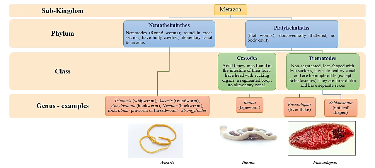
Fig. 1:Taxonomic classification of helminths. There are three main groups of helminths, i.e., Trematodes (flukes), Cestodes (tapeworms), and Nematodes (roundworms). They are classified by their overall external shape and the host organ they inhabit. Helminths develop through egg, larval, and adult stages. The outer covering of the helminths is the cuticle or tegument.
Table 1: Current anthelminthic drugs and their mechanism
| Name of the drug | Structure | Mechanism | Side effects |
|---|---|---|---|
| Fenbendazole | 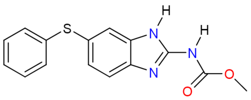 | It binds to tubulin, disrupts the tubulin-microtubule equilibrium, and inhibits polymerization in helminths. It also induces apoptotic cell death of porcine trophectoderm and uterine cells following membrane disruption. It might lead to implantation failure in pigs during early pregnancy.[16,17] | Diarrhea, Loss of appetite, Lethargy |
| Piperazine |  | It is a selective GABA agonist that activates and gates GABA receptors on nematode muscle. It causes hyperpolarization of muscle cells and nerve membranes at the neuromuscular junction leading to parasite immobilization by flaccid paralysis and consequent removal by interfering with the energy metabolism of the nematode.[18] | Blurring vision, clumpiness, joint pain, skin rash, crawling or tingling feeling of the skin, skin rash, or itching. |
| Mebendazole | 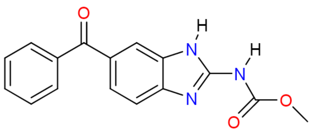 | It inhibits the production of microtubules via binding to the colchicine binding site of β-tubulin and thereby blocking the polymerization of tubulin dimers in the intestinal cells of parasites. Hence, glucose uptake gets interrupted, resulting in immobilization and hindrance of egg production and death of helminth.[19] | Diarrhea, Stomach pain, swelling, nausea, vomiting, loss of appetite. |
| Albendazole | 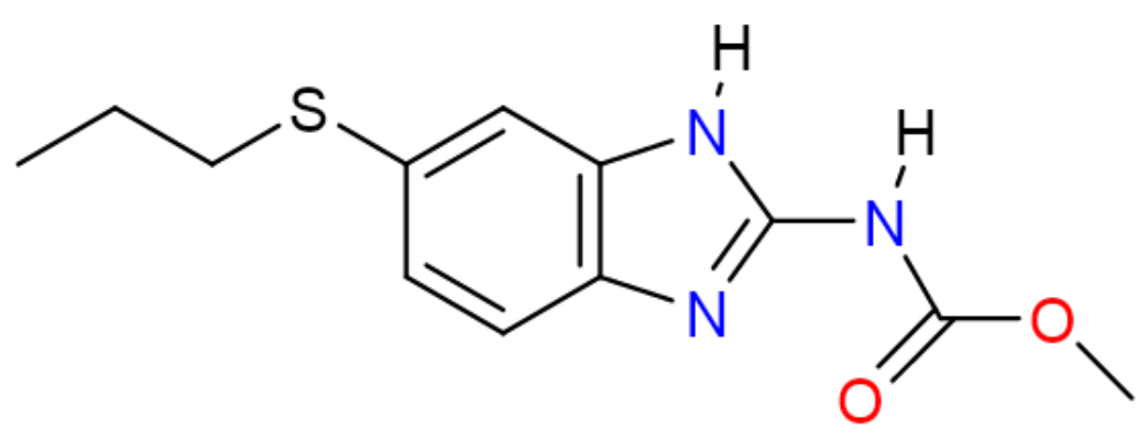 | It binds to the tubulin dimer interface overlapping the binding site of β-tubulin, thereby inhibiting and disrupting microtubule networks.[20] | Headache, neck stiffness, abnormal liver functions, temporary hair loss. |
| Morantel[21] | ![Morantel[21] chemical structure](https://hel1.your-objectstorage.com/hana/grjournals.hanaplatform.com/article-no-26702/table-1Morantel_1789541392507.webp) | It acts as an agonist of the nAChR subtype comprising ACR-26/ACR-27 subunits from H. contortus or Parascarisequorum expressed in X. laevis oocytes. In oocyte expression studies, morantel was seen to cause a non-competitive voltage-sensitive open channel block of the newly characterized A. suum ACR-16 receptor.[22-24] | Dyspnea, Tachypnea, Diarrhea, Tremor, Convulsions, Ataxia. |
| Pyrantel |  | It activates L-subtype nAChR in A. suum.It also causes an open channel block.[24,25] | Nausea, diarrhea, stomach cramps, trouble sleeping, loss of appetite. |
| Levamisole |  | Levamisole opens nematode AChRs that are ligand-gated, non-selective cationchannels.[25] | Agranulocytosis, skin rash, and febrile illness. |
| Ivermectin | 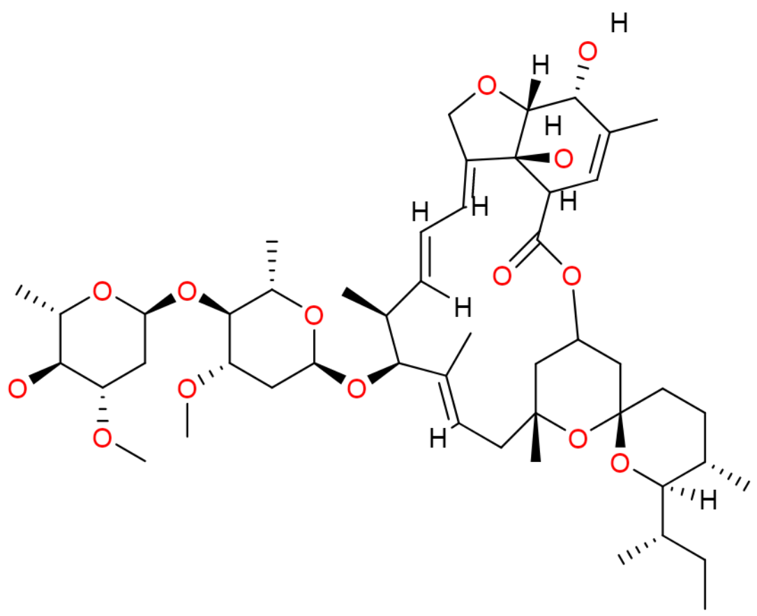 | Ivermectin increases the activity of γ-aminobutyric acid (GABA) receptors or glutamate-gated chloride ion channels (Glu-Cl), which blockades the signal between neurons and muscles.[21,26] | Dizziness, loss of appetite, nausea, diarrhea, weakness. |
by poor bioavailability, limited drug targets, repeated treatment requirements, adverse effects, and, most importantly, the rapid emergence of anthelmintic resistance.[13,14] For example, monepantel, introduced for livestock treatment in 2009, experienced documented resistance in sheep and goats in New Zealand within only three years of its commercial release.[15] Such rapid development of resistance underscores the urgent need for innovative therapeutic approaches. The currently available anthelmintic drugs, their mechanisms of action, therapeutic applications, and major limitations are summarized in Table 1. Nanomedicine has emerged as a promising strategy to overcome many limitations associated with conventional anthelmintic therapy. Nanoparticle-based drug delivery systems can improve drug solubility, stability, and bioavailability, enable
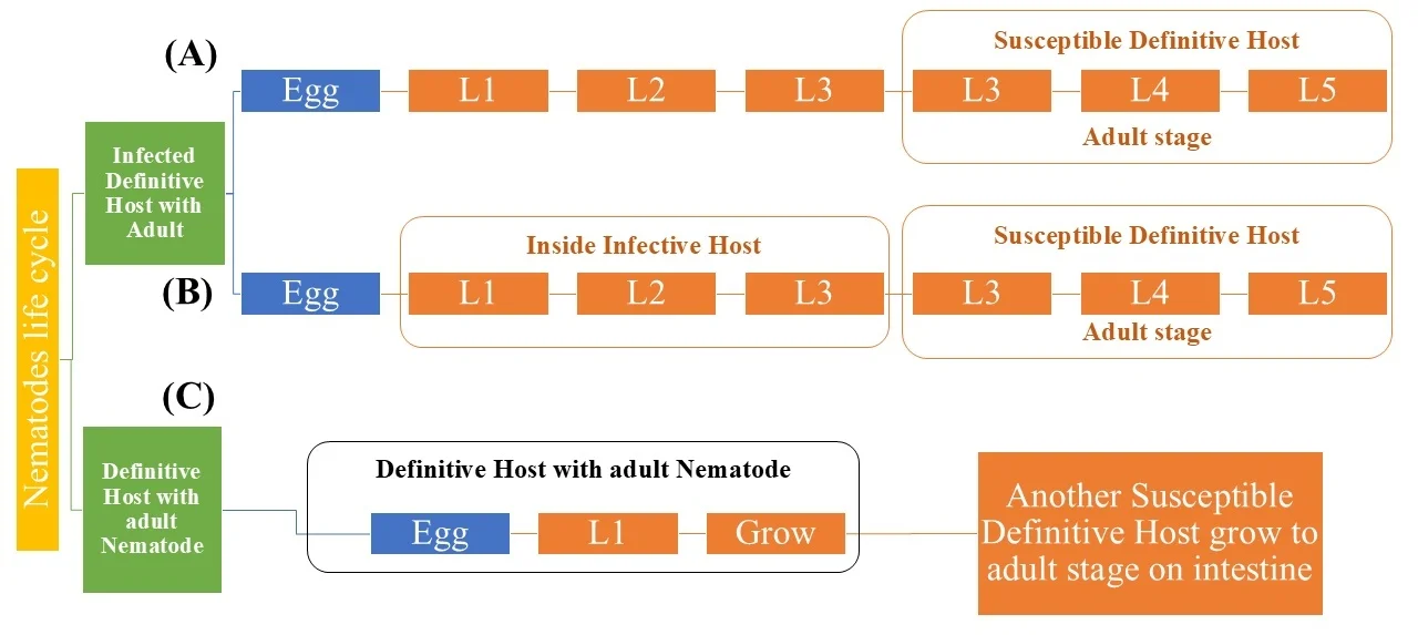
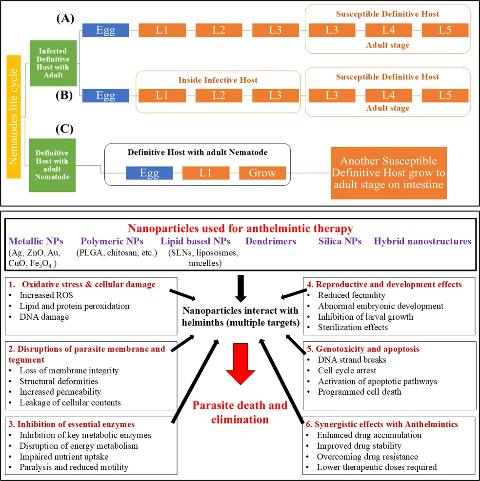
Fig. 2:Life cycle of nematodes (A) direct life cycle (in monogenous nematodes), (B) indirect life cycle (in heterogenous nematodes), (C) indirect life cycle in the self-host (in auto-heterogenous nematodes). There are four stages in a nematode's life cycle: egg, four larval or juvenile stages, and adult. At each juvenile stage, the cuticle sheds, and the nematode increases in size. Nematode's growth depends on the conditions inside the host.
controlled release and targeted delivery, and potentially reduce toxicity and delay the emergence of drug resistance. Furthermore, several nanomaterials possess intrinsic antiparasitic activity and have demonstrated enhanced therapeutic efficacy when used alone or in combination with conventional anthelmintic drugs or herbal formulations. Consequently, nanotechnology represents a promising platform for developing next-generation therapeutics against parasitic worm infections. This review summarizes recent advances in the application of nanomedicine for the management of parasitic worm infections. We discuss different nanoparticle-based delivery systems, their mechanisms of action, therapeutic applications in human and veterinary helminthiasis, their advantages over conventional formulations, current limitations, and prospects for translating these technologies into clinical and veterinary practice.
2. Review of literature
2.1 Helminthic zoonoses
Helminthic zoonoses are parasitic worm infections that are naturally transmitted between animals and humans, representing a major public health and veterinary concern worldwide.[27] Zoonotic diseases account for more than 60% of all human infectious diseases, with helminth parasites contributing substantially to this global burden. Transmission to humans occurs through multiple routes, most commonly via the accidental ingestion of infective eggs or larvae present in contaminated food, water, or soil. For example, Ascaris spp. and Trichuris spp. are primarily transmitted through the fecal–oral route, whereas infective larvae of hookworms and Schistosoma spp. can actively penetrate intact skin. Several helminths possess complex life cycles involving intermediate and definitive hosts, with humans serving as either intermediate or definitive hosts depending on the parasite species (Fig. 2). In contrast, some nematodes, such as roundworms and whipworms, readily infect both humans and animals. Environmental, socioeconomic, and behavioral factors, including sanitation, hygiene, climatic conditions, food preparation practices, dietary habits, and exposure to infected animals or vectors, strongly influence the prevalence and transmission of helminthic zoonoses. Soil-transmitted helminths (STHs) are the most widespread human helminth infections, collectively affecting approximately one-quarter of the global population.[28] The major STHs include Ascaris lumbricoides, Strongyloidesstercoralis, Trichuris trichiura, and the hookworms Ancylostoma duodenale and Necator americanus.[29] In addition to STHs, several foodborne and waterborne helminth infections—including echinococcosis, fascioliasis, taeniasis, cysticercosis, diphyllobothriasis, capillariasis, and ascariasis—remain important but often under-recognized causes of human disease. Their incidence has increased in recent years due to international travel, globalization of the food supply, changing dietary preferences, and the growing consumption of raw or undercooked foods, underscoring the continued need for effective surveillance, prevention, and control strategies.[30]
2.2 Helminth infections and host immune regulation
Helminths have evolved sophisticated mechanisms to evade and manipulate host immune responses, enabling long-term survival within their hosts while minimizing excessive tissue damage. Through millions of years of coevolution, these parasites have established a finely balanced relationship with their hosts by modulating both innate and adaptive immunity, thereby promoting chronic infection and limiting harmful inflammatory responses.[31] A key strategy employed by helminths is the secretion of excretory/secretory (ES) products, including extracellular vesicles (EVs). These vesicles transport a diverse range of bioactive molecules, including proteins, lipids, messenger RNAs, and microRNAs, that regulate host immune signaling pathways and suppress protective immune responses.[32] Growing evidence suggests that EV-mediated communication plays a central role in host–parasite interactions and offers promising targets for the development of novel therapeutics and vaccines.[33] Nevertheless, further studies are required to elucidate the specific functions of individual EV cargo molecules and their interactions with different immune cell populations. Among helminths, Schistosoma spp. provide one of the best-characterized examples of parasite-mediated immune regulation. In addition to causing chronic hepatic, intestinal, urinary, and renal diseases, schistosomes can modulate host immunity in ways that reduce the severity of certain inflammatory and autoimmune disorders. Experimental studies have demonstrated that Schistosoma mansoni infection alleviates allergic airway inflammation, suppresses autoimmune diabetes in mice, and delays disease progression in experimental models of multiple sclerosis.[5] The eggs of S. mansoni not only contribute to disease pathology but also secrete immunomodulatory molecules, including the glycoprotein omega-1, which plays a pivotal role in directing host immune responses toward a regulatory phenotype.[34] Such immune modulation limits excessive inflammation, thereby enhancing host survival while facilitating long-term parasite persistence. Helminth infections are also the leading cause of eosinophilia among travellers, accounting for approximately 30–60% of reported cases, with schistosomiasis representing the predominant diagnosis.[35] Although substantial progress has been made in schistosomiasis control through preventive chemotherapy and public health interventions, implementation remains uneven across endemic regions due to limited healthcare infrastructure and incomplete treatment coverage. Antibody-based assays remain useful for diagnosing travellers and individuals living in low-endemic or post-elimination settings. In contrast, antigen detection and parasite DNA-based assays are emerging as promising tools for field surveillance and disease monitoring.[36] Human hookworms, including Ancylostoma duodenale and Necator americanus, are major causes of iron-deficiency anemia and protein malnutrition, particularly among children. Interestingly, increasing evidence indicates that hookworm infections also possess immunomodulatory properties. Infection with N. americanus has been associated with reduced dust mite sensitization and decreased asthma-related wheezing in children. [37] Likewise, excretory/secretory products from the canine hookworm Ancylostoma caninum suppress allergic airway inflammation in murine models by inducing Th2-associated cytokines, including interleukin-5 (IL-5) and interleukin-13 (IL-13).[38] Furthermore, experimental infection with N. americanus has been shown to reduce intestinal expression of the pro-inflammatory cytokine interferon-γ in patients with autoimmune disorders such as celiac disease.[5] Collectively, these findings demonstrate that, despite their pathogenicity, helminths possess remarkable immunomodulatory properties that may inspire the development of novel immunotherapeutic strategies.
2.3 Current anthelmintic therapy and drug resistance
Despite the enormous global burden of helminth infections, therapeutic options remain limited to only a few classes of anthelmintic drugs. Benzimidazoles (albendazole, mebendazole, and triclabendazole), praziquantel, and macrocyclic lactones such as ivermectin constitute the cornerstone of helminth control in both human and veterinary medicine. The World Health Organization (WHO) recommends albendazole and praziquantel for the treatment of several intestinal helminth infections and for preventive chemotherapy programs, particularly among school-aged children, who represent the principal target population for mass drug administration.[39,40] Each class of anthelmintic drugs exhibits a distinct mechanism of action. Benzimidazoles selectively bind to β-tubulin, disrupting microtubule polymerization, impairing glucose uptake, and ultimately causing energy depletion and the parasite's death.[41] Praziquantel increases the permeability of the parasite's tegument to calcium ions, resulting in sustained muscle contraction, paralysis, and parasite elimination.[42] In contrast, ivermectin targets glutamate-gated chloride ion channels located in the nerve and muscle cells of nematodes, producing membrane hyperpolarization, paralysis, and eventual death of the parasite.[43] Although these drugs have substantially reduced the global burden of helminthiasis, they have several limitations, including limited activity against certain parasite species or developmental stages, poor bioavailability of some compounds (e.g., albendazole), the need for repeated treatment, and the increasing emergence of drug resistance. Treatment failures have now been reported for several human and veterinary helminths, particularly schistosomes and gastrointestinal nematodes, raising concerns regarding the long-term sustainability of existing control programs.[44] Anthelmintic resistance has become especially widespread in livestock, with prevalence exceeding 50% in many regions worldwide, resulting in significant economic losses, reduced animal productivity, and compromised animal welfare.[12] An overview of the major helminth infections affecting humans and animals, together with their first-line therapies and reported resistance, is presented in Table 2. Drug resistance arises through multiple molecular and genetic mechanisms. These include alterations in drug targets that reduce binding affinity; modifications in drug metabolism that enhance detoxification or decrease drug activation; alterations in drug transport, including increased efflux that reduces intracellular drug accumulation; and amplification or overexpression of target genes that reduce drug susceptibility.[12] Additional mechanisms, such as mutations in receptor genes, altered gene expression, and enhanced xenobiotic detoxification pathways, further contribute to the evolution of resistant parasite populations. Recognizing the growing threat of anthelmintic resistance, a Biotechnology and Biological
Table 2: Statistics of parasitic helminth infections in humans and animals and their clinical treatment concerning resistance to anthelmintic drugs
| Common Name | Scientific Name | Disease caused; Symptoms | Human Population infected | Livestock infected | Region affected | Age group infected | Fatality | Treatment | Dosage | Resistance towards drug | Side effects of drug |
|---|---|---|---|---|---|---|---|---|---|---|---|
| Giant roundworm | Ascaris lumbricoides | Ascariasis; Abdominal pain, vomiting, weight loss, fatigue | 807 million to 1.2 billion | NR | Tropical and subtropical regions | 2-10 years | Mostly not fatal | Albendazole | 400 mg PO once [45] | Yes[46] | Headache, fever, nausea, vomiting, stomach pain, temporary hair loss, dizziness[47] |
| Whipworm | Trichuris trichiura | Trichuriasi; Abdominal pain, diarrhea, tiredness | 604-795 million | NR | Tropical regions | 5-15 years | No | Albendazole | 200-400mg | Yes[48] | NR |
| Whipworm | Trichuris trichiura | Trichuriasi; Abdominal pain, diarrhea, tiredness | 604-795 million | NR | Tropical regions | 5-15 years | No | Mebendazole | 100mg twice a day | Yes[48] | Abdominal pain, diarrhea, headache, dizziness[19] |
| Whipworm | Trichuris trichiura | Trichuriasi; Abdominal pain, diarrhea, tiredness | 604-795 million | NR | Tropical regions | 5-15 years | No | Ivermectin | 200 mcg/kg daily [49] | Yes[50] | Loss of appetite, nausea, vomiting, constipation[47] |
| New world hookworm | Necator americanus | Hookworm disease; Abdominal pain, iron deficiency because of chronic blood loss | 576-740 million | NR | South India, Africa, Asia, Australia, America | 5-9 years | No | Albendazole | 400mg once | Yes[51] | Headache, fever, nausea, vomiting, stomach pain, temporary hair loss, dizziness[47] |
| New world hookworm | Necator americanus | Hookworm disease; Abdominal pain, iron deficiency because of chronic blood loss | 576-740 million | NR | South India, Africa, Asia, Australia, America | 5-9 years | No | Mebendazole | 500mg orally once [52] | Yes[53] | Abdominal pain, diarrhea, headache, dizziness[19] |
| Old world hookworm | Ancylostoma duodenale | Ancylostomiasis/ Wakana Syndrome; Rash at the site of larval entry, abdominal pain, iron deficiency because of chronic blood loss | 576-740 million | NR | North India, Middle East, North Africa | < 4 years | No | Albendazole | 400mg once | Yes[54] | Headache, fever, nausea, vomiting, stomach pain, temporary hair loss, dizziness[47] |
| Old world hookworm | Ancylostoma duodenale | Ancylostomiasis/ Wakana Syndrome; Rash at the site of larval entry, abdominal pain, iron deficiency because of chronic blood loss | 576-740 million | NR | North India, Middle East, North Africa | < 4 years | No | Mebendazole | 500mg orally once[52] | Yes[53] | Abdominal pain, diarrhea, headache, dizziness[19] |
| Blood Fluke | Schistosoma species | Schistosomiasis; Fever, chills, cough, muscle aches, abdominal pain, blood in stool or urine. | 236.6 million | NR | Sub-Saharan Africa, South America, -Caribbean, Middle East | 6-15 years | Yes | Praziquantel | 20 mg/kg thrice a day[55] | Yes[56] | Headache, dizziness, stomach pain, nausea, vomiting, joint/muscle pain, sweating[57] |
| River blindness | Onchocercavolvulus | Onchocerciasis; Skin rashes, itching, bumps under the skin, itching of eyes, cataract | 120 million | NR | African Countries | 35-45; primarily males | No | Ivermectin | 150mg/kg once a year | Yes[58] | Loss of appetite, nausea, vomiting, constipation [47] |
| Threadworm | Strongyloidesstercoralis | Strongyloidiasis; Abdominal pain, diarrhea, cough, vomiting, red hives near the anus, weight loss | 3-100 million | NR | Brazil, Central America | 16-45 years | Yes | Stromectol | 200 mcg/kg [59] | Yes[60,61] | Headache, nausea, muscle pain, diarrhea, swelling of hands/ankles/feet, itching[62] |
| Threadworm | Strongyloidesstercoralis | Strongyloidiasis; Abdominal pain, diarrhea, cough, vomiting, red hives near the anus, weight loss | 3-100 million | NR | Brazil, Central America | 16-45 years | Yes | Albendazole | 400 mg twice a day [63] | Yes[61] | Headache, fever, nausea, vomiting, stomach pain, temporary hair loss, dizziness[47] |
| Chinese liver fluke | Clonorchis sinensis | Chlonorchiasis; Indigestion, abdominal pain, diarrhea, constipation, inflammation. | 35 million | NR | Eastern Asia | 40-60 years | Yes | Praziquantel | 25 mg/kg thrice a day [64] | - | Headache, dizziness, stomach pain, nausea, vomiting, joint/muscle pain, sweating[57] |
| Chinese liver fluke | Clonorchis sinensis | Chlonorchiasis; Indigestion, abdominal pain, diarrhea, constipation, inflammation. | 35 million | NR | Eastern Asia | 40-60 years | Yes | Albendazole[65] | 10 mg/kg once a day | - | Headache, fever, nausea, vomiting, stomach pain, temporary hair loss, dizziness[47] |
| Filariae | Roundworms of the Filarioidea type | Lymphatic filariasis; Itchy skin, abdominal pain, muscle pain, swelling under the skin, enlarged liver and spleen, inflammation. | 25 million | NR | Tropical countries | Adults | Rarely fatal | Diethylcarbamazine | Day 1&2: 50 mg PO PC Day 3: 100 mg PO TID Day 4-14: 6 mg/kg/day PO divided TID[66] | Yes[67] | Fever, painful and tender glands in neck, armpits, skin rash |
| Filariae | Roundworms of the Filarioidea type | Lymphatic filariasis; Itchy skin, abdominal pain, muscle pain, swelling under the skin, enlarged liver and spleen, inflammation. | 25 million | NR | Tropical countries | Adults | Rarely fatal | Albendazole; | 400 mg twice a day for 21 days [68] | Yes[67] | Headache, fever, nausea, vomiting, stomach pain, temporary hair loss, dizziness[47] |
| Filariae | Roundworms of the Filarioidea type | Lymphatic filariasis; Itchy skin, abdominal pain, muscle pain, swelling under the skin, enlarged liver and spleen, inflammation. | 25 million | NR | Tropical countries | Adults | Rarely fatal | Ivermectin | Not very effective[68,69] | Yes[67] | Loss of appetite, nausea, vomiting, constipation[47] |
| Southeast Asian liver fluke | Opisthorchis viverrini | Opisthorchiasis, Fever, anorexia, nausea, vomiting, abdominal pain. | 10 million | NR | Southeast Asia | >35 years | Yes | Praziquantel | 75 mg/kg thrice a day for 2 days | Yes[70-73] | Headache, dizziness, stomach pain, nausea, vomiting, joint/muscle pain, sweating[57] |
| Southeast Asian liver fluke | Opisthorchis viverrini | Opisthorchiasis, Fever, anorexia, nausea, vomiting, abdominal pain. | 10 million | NR | Southeast Asia | >35 years | Yes | Albendazole | 100 mg/kg twice daily for 3 days [72,73] | Yes[74] | Headache, fever, nausea, vomiting, stomach pain, temporary hair loss, dizziness[47] |
| Barber’s pole worm | Haemonchuscontortus | Anemia, bottle jaw | 10 million | NR | Sub-Saharan Africa, South Asia | Infants and school-age children | Yes | Ivermectin | 1 mL/5 kg live weight | - Yes, towards amino- acetonitrile derivative class (monepantel) [75] | Loss of appetite, nausea, vomiting, constipation[47] |
| Barber’s pole worm | Haemonchuscontortus | Anemia, bottle jaw | 10 million | NR | Sub-Saharan Africa, South Asia | Infants and school-age children | Yes | Albendazole | 7.5 mg/kg [76] | Headache, fever, nausea, vomiting, stomach pain, temporary hair loss, dizziness[47] | |
| Barber’s pole worm | Haemonchuscontortus | Anemia, bottle jaw | 10 million | NR | Sub-Saharan Africa, South Asia | Infants and school-age children | Yes | Fenbendazole | 5 mg/kg | Salivation, vomiting, diarrhea[77] | |
| Barber’s pole worm | Haemonchuscontortus | Anemia, bottle jaw | 10 million | NR | Sub-Saharan Africa, South Asia | Infants and school-age children | Yes | Moxidectin[76] | 0.2 mg/kg [78] | Increased or decreased WBCs, musculoskeletal pain, headache, fast heart rate, abdominal pain, rash, low BP[79] | |
| Giant Intestinal Fluke | Fasciolopsisbuski | Fasciolopsiasis; Abdominal pain and diarrhea, nausea, vomiting, and fever. | 10 million | NR | Southern and eastern Asia | Females are affected more than males; 10-20 years | Yes | Praziquantel | 75 mg/kg thrice a day for 2 days [80] | - | Headache, dizziness, stomach pain, nausea, vomiting, joint/muscle pain, sweating[57] |
| Giant Intestinal Fluke | Fasciolopsisbuski | Fasciolopsiasis; Abdominal pain and diarrhea, nausea, vomiting, and fever. | 10 million | NR | Southern and eastern Asia | Females are affected more than males; 10-20 years | Yes | Triclabendazole | Two doses of 10 mg/kg 12 hours apart | Yes[70,80] | Nausea, urticaria, vomiting, headache, indigestion, musculoskeletal pain[80] |
| - | Heterophyesheterophyes | Heterophyiasis; Diarrhea and colicky abdominal pain. | 7 million | NR | Middle East, West Europe, Africa | 50-62 years | No | Praziquantel | 75 mg/kg/day in 3 divided doses [81] | - | Headache, dizziness, stomach pain, nausea, vomiting, joint/muscle pain, sweating[57] |
| Cattle Hookworm | Bunostomumphlebotomum | Bunostomosis; Anemia, diarrhea, weight loss, dermatitis | NR | 14.7% in sheep and 29.6% in cattle[82] | Warm and Humid Regions | NR | No | Thiabendazole [83] | 110mg/kg | - | Nausea, vomiting, loss of appetite, upset stomach, diarrhea, dizziness, drowsiness, headache[47] |
| Cattle Hookworm | Bunostomumphlebotomum | Bunostomosis; Anemia, diarrhea, weight loss, dermatitis | NR | 14.7% in sheep and 29.6% in cattle[82] | Warm and Humid Regions | NR | No | Benzimidazole | Yes | Dizziness, anorexia, nausea, and vomiting[84] | |
| Cattle Hookworm | Bunostomumphlebotomum | Bunostomosis; Anemia, diarrhea, weight loss, dermatitis | NR | 14.7% in sheep and 29.6% in cattle[82] | Warm and Humid Regions | NR | No | Levamisole | 10 mg/kg | Yes | Blurred vision, confusion, seizures, lip-smacking, numbness, paranoia, puffing of cheeks, worm-like movement of the tongue[47] |
| Cattle Hookworm | Bunostomumphlebotomum | Bunostomosis; Anemia, diarrhea, weight loss, dermatitis | NR | 14.7% in sheep and 29.6% in cattle[82] | Warm and Humid Regions | NR | No | Ivermectin[85] | Yes | Loss of appetite, nausea, vomiting, constipation [47] | |
| Cattle Hookworm | Bunostomumphlebotomum | Bunostomosis; Anemia, diarrhea, weight loss, dermatitis | NR | 14.7% in sheep and 29.6% in cattle[82] | Warm and Humid Regions | NR | No | Fenbendazole[86] | 10 mg/kg | Salivation, vomiting, diarrhea[77] | |
| Stomach worm | Trichostrongylusaxei | Catarrhal gastritis; weight loss, ulcers | 5 million | NR | NR | NR | Infection in young animals can be fatal | Benzimidazole | 36.7 mg/day | Benzimidazole in sheep, horses [87,88] | Dizziness, anorexia, nausea, and vomiting[84] |
| Common liver fluke | Fasciola hepatica | Fasciolosis; Fever, malaise, abdominal pain, eosinophilia, hepatomegaly. | 2.4 million | NR | Tropical areas | 9-12 years | Yes | Triclabendazole | 10 mg/kg twice a day [80] | Triclabendazole[70] | Nausea, urticaria, vomiting, headache, indigestion, musculoskeletal pain[80] |
*Note: Helminths get transferred to animals and humans through the soil, food, and water. Generally, flu-like symptoms are common, including high temperatures and muscle aches. Symptoms may also include skin rashes, abdominal pain, diarrhea, and cough. Also, inflammation occurs in areas of the body where eggs have lodged, i.e., intestine, eggs, liver, lungs, and even brain. This table includes the activity of antiparasitic drugs, which outlines indications, dosage, adverse effects, and resistance towards these drugs.
NR = Not reported.
Table 3: Potential herbal medicines with anthelmintic properties
| Name of plant | Part of plant used | Traditional Use | Active Components | Acts against | Effect on life cycle | Molecular Mechanism |
|---|---|---|---|---|---|---|
| Artemesia herba-alba | fresh flower and aerial parts | Enteritis and various intestinal disturbances among Bedouins in the Negev desert. | Tannins (Proanthocyanidins) | H. contortus, H. gallinarum, T. colubriformis [89-91] | Decrease in egg hatching, blocking development to the infective larval stage and reduced motility of larvae and adult stage of H. contortus | Tannins interfere with coupled oxidative phosphorylation and block ATP synthesis, Protein damage |
| Punica granatum | peel and root | To treat sore throats, urinary infections, digestive and skin disorders, and expel tapeworms. | Gallic acid | H. contortus (in sheep), A. lumbricoides, E. papillate, A. caliginosa, G. indicus[91-95] | Decrease in egg hatching, ovicidal activity. Decrease in goblet cell numbers at the site of E. papillate infection. | Gallic acid interferes with oxidative coupled phosphorylation and blocks ATP synthesis. Punica granatum prevented the infection-induced loss of GSH and increased the production of NO and MDA. |
| Carica papaya L. | leaves, fruit, and seed | It treats and improves digestive and abdominal disorders. | papain (papaya proteinase I) and Benzyl Isothiocyanate | T. colubriformis, H. polygyrus, P. posthuman, H. polygyrus, H. bakeri[96-101] | Drop-in fecal egg count | The aqueous extract inhibited glucose uptake and depleted glycogen content in the presence of glucose and increased lactate dehydrogenase, thus inhibiting ATP production or accumulation of lactic acid. |
| Allium sativum L. | bulbs | It has preventive characteristics in cardiovascular diseases, regulating blood pressure, lowering blood sugar and cholesterol levels. | acetylcholine | A. galli, H. contortus, F. gigantica, T. canis, A. caninum, H. gallinae (Jeyathilakan et al. 2012)[102-106] | Extract reduced L4 population of H. contortus in values higher than 50% | Acetylcholine suppresses smooth muscle contraction, causing paralysis, and allicin disrupt the glycolytic pathway, and no energy leads to the death of A. galli |
| Cucurbita mexicana | seeds | Seeds are used in Mexico to make palanquetas (sweet,similar to peanut brittle). Jam is made out of its fruit. | Cucurbitacin | A. lumbricoides, H. diminuta, F. gigantica, M. expansa, F. buski[107-109] | Reduction in egg count, inhibition of egg hatching | NH4 concentration dropped due to its oxidation to NO3, causing the elimination of air by water |
| Albizia ferruginea | Stem bark | To treat dysentery, bronchial affections, and pain caused by fever, it is applied externally to sores, pimples, and other skin complaints. | Tannins, saponins, glycosides, alkaloids, and coumarins | P. posthuman, H. contortu [110] | Enhanced disruption of the integrity of the helminth tegument, inhibition of motility. | Reduction in glucose uptake by the worm. Inhibition of homovanilic acid and binds to enzymes. |
| Nigella sativa | seeds | To manage pain during menstruation and treat diabetes and high blood pressure | Thymoquinone, Dithimoquinone, Thymol, and Palmitic acid | S.mansoni, C. cotylophorum[108,111,112] | Inhibition of egg-laying and motility. | Oxidative stress against adult worms and decrease in the activity of antioxidant enzymes (superoxide dismutase and glutathione peroxidase), glutathione reductase, and enzymes of glucose metabolism (hexokinase and glucose-6-phosphate dehydrogenase) |
| Piper longum L. | Fruit and leaves | It is used in India, Malaysia, Singapore, and other South Asian Countries as an analgesic, carminative, and to treat asthma and insomnia. | Piperine, Piperlongumine | A. Lumbricoides, F. giganticum, P. posthuma, [99,113] | Causes 50% inhibition of egg hatching even at the lowest concentration; increase in larval motility; larval migration inhibition (1.95 mg/mL) | Suppression of transfer of sucrose from the stomach to the small intestine, reducing nitrate generation interfering in homeostasis. Also, it inhibits glucose uptake by worms. |
| Ocimum sanctum L. | leaves | To treat bronchitis, bronchial asthma, malaria, diarrhea, skin diseases. | Eugenol, Tannins and flavonoids | C. elegans, A. galli, F. gigantica[114-116] | ethanol extract with 10% concentration and exposure time for 10 hours caused the most mortality. | Tannins increase protein uptake. Flavonoids affect the nervous system leading to neuron degeneration. |
| Azadirachta indica | leaves | Used for leprosy, eye disorders, bloody nose, skin ulcers, etc. | Azadirachtin | F. gigantica, P. cervi, F. hepatica, H. contortus, P. posthuma[117-121] | Deformation of body shape, eggs were blackened. | Depletion of glycogen contents and disturbs calcium homeostasis and Nitrous Oxide activity. |
| Moringa Oleifera | Leaves and seeds | Kings and queens used Moringa oleiferato improve their alertness and maintain healthy skin. | Tannins, Flavonoids, Triterpenoids, Saponins, and Alkaloids | D. medinensis, S. mansoni, B. alexandrina, H. contortus, Trichuris spp., Ostertagia spp. [122-125] | Alterations were seen in the embryonic development of the eggs, where all embryos are at the last stage (pre-hatching). | Increased transaminases (ALT and AST) decrease total proteins, albumin, and globulin concentration and damage digestive cells. |
| Artemisia annua L. | leaves | To treat fever, inflammation, and malaria | Artemisinin, Quercetin | F. hepatica, Schistosoma spp., H. contortus [126-128] | Tegumental alterations and reduction of motor activity of adult worms. | Interference with parasite transport proteins, disruption of mitochondrial function, host immune function modulation, and angiogenesis inhibition. Expression of Hc29 gene causing ROS. ROS dependant neuronal and other tissue damage. |
| Butea monosperma | Seed, root, flower, and leaves | It is an anthropogenic tree of several castes. The use of its gum as an external astringent application is mentioned in “Chakradatta.” | Palasonin and Tannins, phenolics, flavonoids | A. lumbricoides, A. galli, D. caninum, T. canis, H. contortus, C. elegans, Trichostrongylid nematodes, D. caninum[99,129] | Peptide inhibitor affects the growth and development physiology of the helminths. | Affects the activity of gut proteases. |
| Terminalia arjuna | Leaf, bud, root, seed, and stem, bark | In traditional ayurvedic medicine, Terminalia arjuna balances three “humor”: Kapha, Pitta, and Vata. | Condensed tannin and ellagic acid | Eggs, larvae, and adults of H. contortus, P. posthuma[130-133] | Condensed tannins cause larval starvation. | Decrease in gastrointestinal metabolism by inhibition of oxidative phosphorylation. Also, inhibition in key enzymes, i.e., AChE and AcPase activities. |
| Cymbopogon martini | Whole plant | The essential oil has applications in aromatherapy. Therapeutic properties include antiseptic, antiviral, and antibacterial. | Geraniol | C. Elegans, P. posthuma[134,135] | Inhibiting egg hatching and larval development. | Geraniol triggers enzymatic activity in the worms. |
| Mentha cordifolia | Leaf | Used in cooking, cosmetics, treatment of gastrointestinal disorders. | Glucosides, β-Sitosterols | A. suum[136] | Reduction in egg count. | β-Sitosterols trigger apoptosis, higher caspase activity. It minimizes the number of squirms caused by acetic acid. |
| Calotropis Procera | Leaves | Used for diarrhea, stomatic, sinus fistula, and skin disease. | Anthocyanins, Triterpenoids, alkaloids | H. contortus, P. posthuma[137,138] | Disorganization of the cuticle, muscle layer degradation. Reduction in egg count. | Cysteine proteases cause changes in the cuticle and inhibit the motility of the worms. |
| Aloe ferox | Leaf | Yellow bitter sap is used as a laxative, and white gel is used in health drinks and skincare products. | Phenol, , Alkaloids, and Flavonoids | Trichuris spp., H. contortus, Trichostrongylus, Teladorsagia nematodes, H. gallinarum[139-141] | Affects egg-laying ability of female adult worms | Blocks glucose uptake. |
| Chelidonium majus | Upper part (Stem, leaves) | To treat blood stasis, relieve pain, to treat jaundice | Chelerythrine; Chelidonine | T. canis, D. intermedius[142] | Mortality after 24 hours | Chelerythrine exhibit cytotoxic properties against normal cells, exerting apoptosis-inducing pathways. |
| Macleaya spp. | Upper part (Stem) | Macleaya cordata is a traditional antiviral, anti-inflammatory, and insecticidal herb medicine | 6-Methoxydihydrosanguinarine | T. canis, T. spiralis, D. intermedius[142-144] | Mortality after 24 hours; Decrease in rate of eggs hatching | Sanguinarine increases intestinal goblet cell hyperplasia. |
| Onobrychis viciifolia | peel | For stabilizing soils | Rutin, Nicotiflorin, Narcissin, Condensed Tannin | H. contortus, O. ostertagi, C. oncophora, T. colubriformis[145,146] | Inhibition of migration of L3 worms and larval feedings. | Tannins interfere with coupled oxidative phosphorylation and block ATP synthesis. |
| Cissampelos capensis | Upper part | For treatment of intermittent fever, heatstroke | (S)-Dicentrine, 63(S)- Neolitsine | H. contortus, P. posthuma[147,148] | Inhibition of larval development. | An increase in chloride ion conductance of worm muscle membrane produces hyperpolarization and reduced excitability leading to flaccid paralysis. |
| Acacia oxyphylla | Stem bark | Acacia oxyphylla has been traditionally used by natives of Mizoram against intestinal worm infections. | 12-Amino-7,17-dioxo-2-oxa-8,16-diazatricyclo [14.2.2.2 3,6] tetraicosa-1 (20),3,5,18,21,23-hexaene-12-carboxylic acid (F5-2d) | A. galli, R. echinobothrida [149-151] | Cysteine proteinases cause wrinkles and folds of the cuticle, followed by blistering and gradual digestion of the cuticle. | AcPase and AlkPase activity decreased. |
| Eryngium foetidum | Whole plant | Used in tropical regions for burns, earache, fever, hypertension, constipation, fits, asthma | Eryngial | S. stercoralis, Paramphistomum spp., P. posthuman, T. tubifex [152,153] | Larval mortality after 24 hours. | Inhibition of α-glucosidases. |
| Cinnamomum verum | Bark | To treat headache, chills, abdominal pain, cough, chest tightness, diarrhea | Trans-Cinnamaldehyde | A. suum, T. suis, O. dentatum, A. galli, P. posthuma, H. contortus[154,155] | Larval mortality after 12 hours. | Trans-cinnamaldehyde, an α, β unsaturated aldehyde, reacts with nitrogen groups, interferes with key enzymes, and can cause cell lysis. |
| Dichapetalum filicaule | Whole roots | Used as a food additive; solidifier and control release agent in pharmaceuticals; resin lubricant. | Dichapetalin A, Glycerol monostearate | N. americanus, S. haematobium[156] | Inhibition of egg hatching | The opening of glutamate-gated chloride channels, increasing the flow of chloride ions and leading to defects in neurotransmission and flaccid paralysis |
| Thymus vulgaris | Leaves | Used as herbal tea, stimulant, Antiflatulent, cough depressant, treatment of a common cold. | Thymol, Thymol acetate | H. contortus, E. granulosus[157,158] | Effective against three stages of H. contortus: Egg hatching, larval development, and adult stages. | Enzyme (GGT) activity decreased. |
| Melaleuca alternifolia | Leaves | Antiseptic and anti-inflammatory properties | Terpinen-4-ol | H. contortus, A. simplex[159,160] | TTO inhibits in vitro egg hatch, larval migration | terp-4-ol was able to prevent increased levels of AST and ALT. |
| Ajania nubigena | Aerial part | Have antimalarial properties | Luteolin, (3R,6R)- Linalool oxide aceta | T. muris, S. mansoni[161] | Luteolin induces tegumental damage to S. mansoni and affects the cuticle, bacillary bands, and bacillary glands of T. muris | Compounds Interfere with coupled oxidative phosphorylation. |
| Mundulea sericea | Upper part | Zulus use the leaves as an emetic to treat poisoning in both people and dogs | Deguelin | H. contortus[142,162] | Caused mortality but at lower rates. | Deguelin exerts a toxic effect on nematodes as a modulator of oxidative phosphorylation. |
| Ruta chalepensis | Air-dried aerial parts | Used as an anti-inflammatory, analgesic, antipyretic agent | 2-Decanone, 2-Nonanone, 2-Undecanone | Teladorsagia spp., H. contortus, and Trichostrongylus spp., O. trigotephras [142,163,164] | Inhibition of egg hatching and larvae motility. | Oxidative Stress |
| Gliricidia sepium | leaves | Used for live fencing, fodder, firewood, green manure, intercropping, and rat poison | 2H-Chromen-2-one | C. punctata[165] | Electron density alterations and fractures in eggshell layers. | Anti-exsheathment activity. |
| Avena sativa | Oat seeds | To treat nervous exhaustion, insomnia, and weakness of nerves. | AveB and 26DGAveB | H. bakeri[166] | AveB and 26DGAve B leads to abnormalities in L1 larvae, inhibits egg hatching | Altered embryogenesis in the egg; overexpression of CED-9 preventing apoptosis; ATP inhibition. |
| Tagetes filifolia | Aerial Parts | As a food flavoring, a tea, and a diuretic | Chlorogenic acid | H. contortus[153,167] | Inhibits egg hatching | ROS-induced cytotoxicity. |
| Acacia cochliacantha | Leaves | As an antiseptic, demulcent, purgative, and effective tonic in diabetes mellitus | Caffeoyl and coumaroyl derivatives: Caffeic acid, p-coumaric acid, Ferulic acid, Methyl caffeate, Methyl p-coumarate, Methyl ferulate, Quercetin | H. contortus[168] | Ovicidal and larvicidal activity | ROS dependant neuronal and other tissue damage. |
| Persea americana | Seeds | Leaves used against dysentery, coughs, high blood pressure, liver problems, and gout | Epicatechin, rutin, chlorogenic acid, quercetin | H. contortus[142,169] | Inhibits L3 motility. | ROS-induced cytotoxicity. |
| Ficus benjamina | Latex | To treat skin disorders, inflammation, piles, vomiting, leprosy, malaria, nose diseases. | CM-cellulose, a cysteine protease (FbP) | H. contortus[170] | Degradation of the cuticle of adult nematodes. | Hydrolysis of peptide bonds of the protein. |
| Baccharis conferta | Aerial parts | Forage for farm animals. | Isokaempferide | H. contortus[171] | Inhibits egg hatching. | Isokaempferide crosses the eggshell layer without breaking it and attaches itself to the embryo, causing its death. |
| Senegalia gaumeri | Leaves | - | p-Coumaric acid | H. contortus[172] | Ovicidal activity; larvae failing eclosion | Ovicidal activity; larvae failing eclosion |
| Alectryon oleifolius | Whole plant | - | Procyanidin A2 | Cyathostomins[173] | Procyanidin A2 completely inhibits development from egg to third larval stage. It also inhibits the larval migration. | Triggers apoptosis. |
| Brazilian Red Propolis | Whole plant | Mummification rituals, and more recently in the industry of food and beverages, cosmetics. | Need to be determined in future | T. cati, S. mansoni[174,175] | Larvicidal activity | Mechanism not known. |
| Caesalpinia coriaria | Fruits | As an antiperiodic and for dressing sores | Gallic acid | Gastrointestinal nematodes (Cooperia spp., Haemonchus spp., Ostertagiaspp., Trichostrongylus spp., and Oesophagostomum spp.)[176,177] | Inhibits egg hatching | Comounds interact with eggshell proteins. |
| Andrographis paniculata | Leaves | Treatment of cancer, diabetes, high blood pressure, ulcers, bronchitis, influenza, dysentery, and malaria. | Andrographolide | A. duodenale, H. contortus[178,179] | Andrographolide inhibits egg hatching and larval motility. | Mechanism not known |
| Cuminum cyminum | seeds | Skin infection, fever, vomiting, nausea, anticancer, antidiabetic, neuroprotection, and anti-inflammation | Cuminaldehyde | H. contortus[78] | Induces adult worms and larvae L3 mortality and inhibits egg hatching. | Oxidative stress-mediated cell and tissue damage (including ROS and NOS). |
*Note: This table discusses the herbal plants used against different helminth infections, along with their active components, structure, and mode of action.
Sciences Research Council (BBSRC)-sponsored workshop identified four major research priorities: the development of improved diagnostic tools for resistance detection, a better understanding of drug mechanisms and resistance pathways, deeper insights into parasite biology, and the establishment of predictive decision-support systems for sustainable parasite control. These priorities emphasize the urgent need for integrated management strategies that extend beyond reliance on conventional chemotherapy. Among livestock parasites, gastrointestinal nematodes (GINs) remain one of the greatest constraints on profitable animal production worldwide. The widespread emergence of resistant parasite populations, together with the limited number of available drug classes and the high cost of developing new anthelmintics, has stimulated interest in alternative control strategies. Plant-derived anthelmintics, widely used in ethnoveterinary medicine, have demonstrated promising antiparasitic activity in numerous experimental studies (Table 3). However, their efficacy is often influenced by variability in phytochemical composition, dosage, host nutrition, and potential anti-nutritional effects. Consequently, standardized evaluation methods and integrated approaches are essential for maximizing their therapeutic benefits while minimizing adverse effects. These limitations have accelerated interest in nanotechnology-based drug delivery systems, which offer new opportunities to improve the efficacy, bioavailability, and targeted delivery of existing anthelmintic agents.
2.4 Nanotechnological approaches for eliminating helminthic diseases
The rapid emergence of anthelmintic resistance, coupled with the limited number of available drug classes, has accelerated the search for innovative therapeutic strategies. Nanotechnology has emerged as a promising platform for improving the diagnosis, prevention, and treatment of parasitic diseases by enhancing drug delivery and therapeutic efficacy.[180] Nanomaterials, typically ranging from 1 to 100 nm in size, are composed of metallic, non-metallic, or hybrid materials whose physicochemical properties can be tailored through controlled synthesis and surface functionalization.[181] Polymer coatings are commonly employed to improve biocompatibility, prolong circulation time, and enable selective targeting of biological molecules.[182] The size, morphology, and surface characteristics of nanoparticles are influenced by synthesis parameters, including precursor composition, surfactants, reaction temperature, solvent system, and reactant concentrations (Table 4).[183] Conventional anthelmintic drugs primarily target parasite neuromuscular function or essential metabolic pathways, leading to paralysis, starvation, and eventual elimination of the parasite.[184] However, their effectiveness is often compromised by poor aqueous solubility, limited bioavailability, nonspecific tissue distribution, rapid clearance, and the increasing prevalence of drug-resistant parasite populations.[185] Nanotechnology offers an attractive solution to these limitations by improving drug stability, controlled release, cellular uptake, and site-specific delivery while reducing systemic toxicity. Consequently, nanoparticle-based drug delivery systems have gained considerable attention for enhancing the efficacy of existing anthelmintic agents rather than relying solely on the discovery of entirely new drugs.
An additional advantage of nanoformulation is its potential to mitigate transporter-mediated mechanisms of anthelmintic resistance. In resistant parasites, increased expression or activity of drug-efflux transporters can reduce the intracellular accumulation of anthelmintic compounds, thereby decreasing their access to molecular targets. Encapsulation within polymeric or lipid-based nanocarriers, such as PLGA nanoparticles, solid lipid nanoparticles (SLNs), and liposomes, can alter the physicochemical form and cellular trafficking of the drug, reducing its immediate exposure to membrane-associated efflux systems. Following uptake of the nanocarrier via endocytic or other membrane-associated processes, the encapsulated drug can be released intracellularly in a controlled manner, providing sustained drug exposure and potentially increasing the fraction of drug that reaches its intracellular or membrane-associated targets. In addition, modulation of particle size, surface charge, lipid composition, and surface functionalization can influence membrane interaction, cellular uptake, and intracellular trafficking, thereby providing delivery routes that differ from those of the freely dissolved drug. Thus, nano-delivery may partially circumvent rapid efflux by modifying the route, kinetics, and intracellular availability of the anthelmintic rather than directly inhibiting the resistance mechanism itself. This distinction is important because a nanocarrier's ability to overcome efflux-mediated resistance likely depends on the physicochemical properties of the carrier, the anthelmintic compound, the relevant transporter, and the parasite species. Consequently, nanocarriers may complement conventional resistance-management strategies by improving drug exposure at the parasite target while reducing the extent to which transporter-mediated drug extrusion limits therapeutic efficacy. Nanomedicine applies engineered nanomaterials for disease diagnosis, prevention, and therapy.[186-189] Although initially developed for applications such as oncology and infectious diseases, advances in nanomedicine have provided valuable strategies to improve antiparasitic chemotherapy. Compared with conventional formulations, nanocarriers can increase the solubility of hydrophobic drugs, protect active compounds from premature degradation, prolong circulation time, and facilitate targeted delivery to infected tissues. These advantages may reduce dosing frequency, improve therapeutic outcomes, and minimize adverse effects, making nanomedicine an attractive approach for both human and veterinary helminthiasis.[190]
A wide variety of nanocarriers have been investigated for anthelmintic drug delivery, including polymeric nanoparticles, lipid-based nanoparticles, dendrimers, metallic nanoparticles, micelles, and liposomes (Table 4).
Table 4. Anthelmintic activity of various nanoparticles and their mode of action
| Nanoparticles | Size and Shape | Surface Protection | Synthesis | Tested Against | Dose | Mechanism |
|---|---|---|---|---|---|---|
| Ag | 78.5 to 100 nm; mostly circular | Polyaniline | 100 mL aq. Solution of 1.0 x 10⁻³ M silver nitrate mixed with 300 mL aq. Solution of 2.0 x 10⁻³ M sodium borohydride. Ag ions were then reduced and formed NPs. Ag NPs were then mixed with Momordica charantia aq. extract. | Adult earthworms P. posthuma | 50 mg/ml | +ve charge on Ag attracts -vely charged cell membrane of the parasites. The phytochemicals (M. charantia) attach with glycoprotein on the parasite cuticle and cause death.[191,192] |
| Ag | 17 ± 9 nm; spherical | Aqueous extract of L. parasiticum (ALE) (pH 7.0) | 240 µM AgNO3 wasadded to 5000 µl of ALE. | H. contortus | IC50 value of 144.4 ± 3.1 nM at 48h of exposure | LAgNPs generated oxidative stress and mediated physical damage in worm tissue, leading to a sharp increase in stress-responsive enzyme activity.[193] |
| Ag | 14.51±3.25 nm; spherical | NR | 100 ml AgNO3 added to fungal filtrate (D. flagrans) in the concentration of 1:50 | A. caninum | IC50 value of AgNPs (chemical synthesis) 228.2 ± 6.7 µg/mL ; and AgNPs (D. flagrans) 43.4±6.8 µg/mL | AgNPs penetrate cuticles of larvae, causing changes in tegmentum and consequently the death of the nematode.[194] |
| Ag | 35 ± 15 nm (ARGOVIT); 1-3 nm (UTSA) | NR | ARGOVIT AgNP solution- 200 mg/mL (20%) of polyvinyl-pyrrolidone (PVP); UTSA solution | Cichlidogyrus spp. of Monogenean parasites of fish | 36 and 6μg/L UTSA AgNPs | Swelling, loss of grooves, and disruption of the parasite’s tegument.[195] |
| Ag | 10-15 nm; spherical | NR | 0.017 g AgNO3 dissolved in deionized water and NaOH (0.01 M) | B. malayi | IC50 dose of AgNPs 5 μM | AgNPs caused a decrease in reduced glutathione (GSH) levels and an increase in both protein carbonylation and nitric oxide (NO) levels indicating oxidative stress and nitrosative stress.[196] |
| Ag | ∼8 nm; quasi-spherical | Tribulus terrestrisextract | 1 ml of ethanolic seed extract of Tribulus terrestris was added to 5 ml of 1 mM silver nitrate (AgNO3) solution prepared in deionized double distilled water | G. explanatum | 20 to 50μg/ml of AgNPs | Antioxidant system and detoxification activity of worms get disrupted along with pronounced DNA damage accompanied by an increase in lipid peroxidation and protein carbonylation levels (oxidative stress markers)[197] |
| Ag | 80.42 nm; spherical | Penicillium aculeatum | P. aculeatum culture was used, and 3 mM AgNO3 | E. granulosus | 0.15 mg/mL | AgNPs show promising scolicidal activity.[198] |
| Ag | 30 nm | AgNP suspension at a 1mg/mL concentration in deionized water was prepared by probe sonication for 30 min on ice, and serial 1:2 dilutions were made in deionized water. | S. japonicum | 125 μg/mL | AgNPs induced cercarial tail-shredding, agitated behavior, and a decrease in cercarial secretion.[199] | |
| ZnO NPs | ~ 16.7 nm; hexagonal | NR | 0 ml of freshly extracted egg white (5 mg/mL) was mixed with a 70 ml aqueous solution of 0.25 M zinc acetate. | Ascaridid Nematode; P. equorum | 400 mg/L ZnO NPs | ZnONPs increase levels of ALT, AST, and ALP (muscle damage, gonad injury); total protein content enhancement (worm cellular dysfunction), increase in MDA level (cell membrane damage), depletion in GST and GSH activities, NO increase.[200] |
| ZnO NPs | 16.7 nm; wurtzite (hexagonal) | Egg albumin | Freshly extracted 30 ml egg albumin (5 mg/mL) was mixed into 70 ml aqueous 0.25M zinc acetate solution; stirred 20min and precipitated by adding NH3 at ~pH 7.0 and centrifuged at 5000 rpm for 10 min. | G. explanatum (Parasite of Indian Water Buffalo) | 240μg /ml or 0.012% ZnO NPs | Production of reactive oxygen species that target macromolecules including nucleic acid, proteins, and lipids involved in cellular functions.[201] |
| ZnO NPs | 20–30 nm; spherical | phosphate-buffered saline (PBS) | stock suspension of the ZnO-NPs prepared in PBS (pH = 7.4) by suspending 10mg ZnO-NPs per ml solution | H. contortus | 16 ppm | Induction of oxidative/ nitrosative stress and DNA damage.[202] |
| ZnO NPs | 20–30 nm; spherical | phosphate-buffered saline (PBS) | stock suspension of the ZnO-NPs prepared in PBS (pH = 7.4) by suspending 10mg ZnO-NPs per ml solution | T. circumcincta [192] | 12-16 ppm | Induction of oxidative/ nitrosative stress and DNA damage.[202] |
| CDS NPs (Cadmium sulfide, CdS-G, and CdS-M) | 6.9 nm (CdS-G) and 6.7 nm (CdS-M); crystallite | Hydroxyl radicals | 8gm of cadmium chloride was dissolved in 100ml of deionized water, followed by the addition of 100ml aq. solution of 1M maltose/glucose and 1M sodium sulfide. | P. posthuma | CdS-M nanoparticles show early paralysis and short lethal times in comparison to CdS-G[203] | - |
| Gold NPs | 6 to 18 nm; spherical | N. oryzae | gold chloride (HAuCl4) was added to the culture filtrate | Raillietina echinobothrida & Fasciolopsis buski | 1.0 mg/ml | Tegumental damage and disorientation of microtriches, spines, and scales.[204,205] |
| TiO2 and ZnO NPs | 11 nm (TiO2 NPs) and 21 nm (ZnO NPs) | NR | NR | C. elegans | 172 mg/ml (TiO2 NPs) 1.125 mg/ ml (ZnO NPs) | ROS generation, leading to oxidative stress-mediated inflammation, genotoxicity, and cytotoxicity[206] |
| Rhodamine B-labeledmelatonin (Mel)-loaded lipid-core nanocapsules (LNC) | 201-220 nm | NR | At 38°C, the organic phase (63 mL acetone, 0.025 g melatonin, 0.092 g sorbitan monostearate, 0.403 mL caprylic/capric triglyceride, and 0.250 g PCL) injected in aq. phase (50 mL water and 0.1925 g PS80). After 10 minutes, the organic solvent was eliminated, and water partially evaporated under reduced pressure (at 40°C) to reach 23–24 mL of white opaque liquid product. | C. elegans [207] | 23.70×10¹² to 118.50×10¹² particles/mL | NR |
| ZnO& L-carnitine | 25–40 nm, spherical shape | NR | A stock solution was prepared by dissolving 0.756 mg of ZnO nanoparticles in 5.4 ml (0.9%) normal saline and stored at 5 °C for daily injection | S. mansoni | 5.6 mg/kg (ZnO NPs) and 500 mg/kg (L-carnitine NPs) | Decrease in brain oxidative stress parameter[208] |
| MgO NPs | NR | NR | Magnesium nitrate was dissolved in distilled water, and NaOH was added to form magnesium hydroxide, which was washed with distilled water and dried at 100°C. | M. incognita | 100 ppm | Deformation of cuticle surface due to exudation of body fluids[209] |
| chitosan-albendazole (ChABZ) and chitosan-praziquantel (ChPZQ) nanoparticles | NR | NR | ABZ powders were dissolved in glacial acetic acid, and PZQ was dissolved in 96% ethanol and were then added to the prepared chitosan solution separately. Then, an aqueous TPP solution (1.2 mg/ml) was added. | E. granulosus (Cystic echinococcosis) | Combination of ChABZ and ChPZQ is more effective (5 and 10 µg/ml) [210] | NR |
| Gold (Au) and silver (Ag)NPs | NR | NR | NR | B.alexandrina & S. mansoni[211] | 30 μg/ml Ag and 160 μg/ml Au NPs | NR |
| Silica NPs | 12 and 105 nm | Rhodamine | NR | C. elegans | NR | Disruption of OPT-2/PEP-2- dependent trafficking of nutrient peptides in the intestinal epithelium.[212] |
| Barium ferrite NPs | < 100 nm; hexagonal | NR | NR | C. elegans | 500 µg/mL | Inhibits reproduction and enhance ROS production[213] |
| Thymoquinone-loaded Albumin NPs | 271.3 nm | Thymoquinone | NR | Planarian platyhelminths [214] | 50 µg/mL | NR |
| Phytol loaded PLGA NPs | NR | Phytol | NR | C. elegans | NR | Phytol-PLGANPs suppresses neuronal Aβ expression induced defect in chemotaxis behaviour[215] |
| Cadmium- Selenium NPs | NR | NR | NR | C. elegans [216] | NR | NR |
| CuO NPs | NR | NR | NR | C. elegans | NR | Degeneration of dopaminergic neurons[217] |
| ZnO and FeO NPs | 20-30 nm (ZnO NPs) and 20-40 nm (FeO NPs) | NR | A stock solution of both NPs prepared by suspending them at a concentration of 10 mg/ml using a sonicator probe at 30 W for 10 min, while the working solutions of 0.004, 0.008, and 0.012% (w/v) were prepared with Ringer solution | T. vitulorum | 0.012% (w/v) | decrease worm’s mobility, increase mortality rate as well as elevate MDA and NO content. Oxidative/nitrosative levels also elevated[218] |
| Selenium NPs | 20-150 nm | Streptomyces spp. (M10A65) | Streptomyces spp. (M10A65) culture mixed with 100 mL of ISP2 medium. Inoculated flask incubated. Then, actinobacterial biomass was separated from the medium. | P. posthuma[219] | NR | NR |
| FeO NPs (SPIONs) | 0.35 mmol iron acetylacetonate was dissolved in 4.5 mL benzyl alcohol. | C. elegans | NR | Oxidative stress[220] | ||
| Solid lipid nanoparticles (SLNs) | 165± 103 nm | Albendazole | Double emulsion technique, where the hot ethanolic extraction of beeswax was done using the soxhlet apparatus at 78 oC, followed by drying till all ethanol gets evaporated and then dissolving it in chloroform (50mg/ml). Abz (100 mg/ml) was then added with 2% aqueous Poloxamer 407 (w/v). | H. contortus | 20 µM | Reactive Oxygen Species stress[221] |
| Curcumin loaded NPs | NR | NR | Curcumin-loaded PLGA poly (lactic-co-glycolic acid) nanospheres by the nanoprecipitation technique | S. mansoni[222] | 50-100 µM | Reduction in egg count, increase in liver granuloma volume and collagen content, enhancement in catalase activity, and restoration of enzymatic activities of normal liver cells.[223,224] |
| Curcumin loaded NPs | NR | NR | NR | C. parvum | 50 µM[225] | NR |
| Curcumin loaded NPs | NR | NR | NR | T. gondii[226] | 100 and 200 mg/kg/day for seven days | |
| Curcumin loaded NPs | NR | NR | NR | I. multifiliis[227] | NR | NR |
| Curcumin loaded NPs | NR | NR | NR | A. galli[228] | 100 mg/ml | NR |
*Note: In this table, we have discussed multiple nanoparticles tested against different helminths and their mechanisms. The most-reported methods of action of nanoparticles are antimicrobial activity, ROS-induced cytotoxicity, genotoxicity, plant growth promotion, etc. It has been successfully demonstrated that the actions of nanoparticles are governed by their size, shape, dose, and concentration.
NR = Not reported.
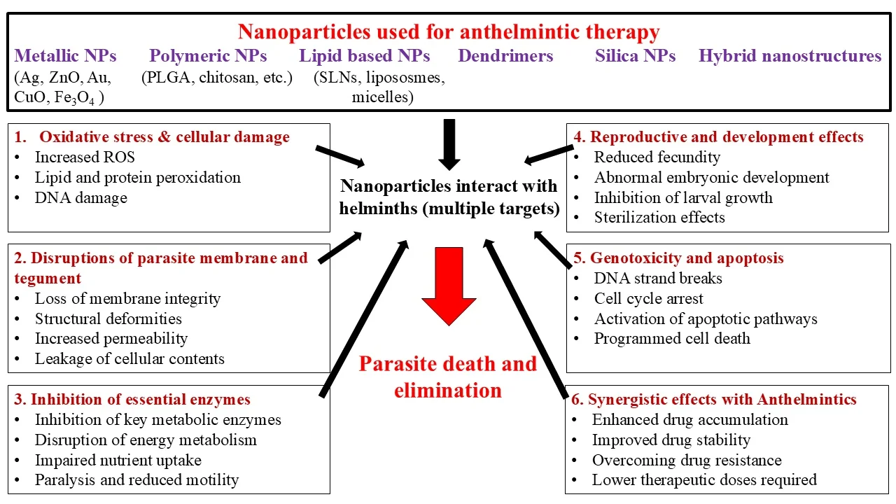
Fig. 3:Graphical representation of Anthelmintic activity of various nanoparticles and their mode of action.
Poly(lactic-co-glycolic acid) (PLGA) remains one of the most extensively studied biodegradable polymers because of its excellent biocompatibility and controlled drug-release properties.[229] Encapsulation of praziquantel within PLGA nanoparticles has been shown to improve drug stability and modify release kinetics. In contrast, solid lipid nanoparticles (SLNs) have demonstrated enhanced oral bioavailability of poorly water-soluble drugs.[230] Likewise, liposomal formulations improve drug encapsulation efficiency, prolong systemic circulation, and promote targeted drug delivery, thereby increasing therapeutic efficacy while reducing systemic toxicity.[231] In addition to functioning as drug carriers, several nanoparticles exhibit intrinsic antiparasitic activity. Metallic nanoparticles, polymeric nanomaterials, and certain hybrid nanostructures have been reported to induce reactive oxygen species (ROS)-mediated oxidative stress, disrupt parasite membranes, impair reproductive capacity, and cause genotoxic damage, ultimately leading to parasite death (Fig. 3). When combined with conventional anthelmintic drugs, these nanomaterials often produce synergistic effects, enabling lower therapeutic doses and improving treatment efficacy against resistant parasites. However, the promising activity reported in experimental studies does not necessarily translate into effective in vivo therapy. Metallic nanoparticles may exert strong antiparasitic effects via oxidative stress, membrane disruption, or genotoxicity, but these mechanisms can also cause nonspecific toxicity in host tissues, thereby narrowing the therapeutic window. Moreover, their biological activity is strongly influenced by particle size, surface characteristics, dose, and aggregation state, meaning that effects observed at relatively high concentrations in isolated parasites may not be reproducible following administration in vivo. The studies summarized in Table 4 also predominantly involve experimental models such as Caenorhabditis elegans, Pheretima posthuma, or isolated parasite stages, which limits direct extrapolation to infected mammalian or livestock hosts. Polymeric and lipid-based nanocarriers may provide greater opportunities for controlled drug delivery, but they also face important barriers to in vivo translation. Although PLGA nanoparticles, SLNs, and chitosan-based systems can improve drug solubility, stability, and release characteristics, their in vivo performance may be compromised by protein corona formation, aggregation, uptake by the mononuclear phagocyte system, premature clearance, and inadequate delivery to the parasite site. Thus, the promising activity of the PLGA-, SLN-, and chitosan-based formulations summarized in Table 4 should be interpreted alongside the need for pharmacokinetic, biodistribution, toxicity, and infected-host studies before therapeutic efficacy can be established.
Despite these promising advances, several challenges continue to limit the clinical translation of nanomedicine. Following administration, nanoparticles encounter numerous biological barriers that influence their absorption, biodistribution, metabolism, and excretion (ADME). Interactions with plasma proteins lead to the formation of a protein corona, which alters nanoparticle stability, circulation time, and tissue distribution. Most nanoparticles are ultimately cleared by the mononuclear phagocyte system, particularly in the liver and spleen, thereby limiting their accumulation at target sites.[232,233] Consequently, optimizing nanoparticle physicochemical properties remains a major focus of current research. These ADME challenges are particularly important for veterinary applications involving food-producing animals. Unlike conventional small-molecule anthelmintics, nanoformulations may exhibit altered tissue distribution, prolonged retention, and formulation-dependent drug release, which can influence the depletion of active compounds and nanoparticle-associated components from edible tissues. Consequently, nanoparticles or their associated drug residues may potentially persist in tissues such as muscle, liver, kidney, and fat and, in lactating animals, may also be transferred into milk, raising concerns regarding consumer exposure. However, the extent of such accumulation and persistence is highly dependent on nanoparticle composition, particle size, surface characteristics, route of administration, dose, and the physicochemical properties of the encapsulated drug. Therefore, residue depletion cannot be reliably extrapolated from conventional formulations of the same anthelmintic. Dedicated pharmacokinetic and tissue-residue studies in relevant livestock species are required to characterize absorption, distribution, transformation, and elimination and to determine the persistence of both the active pharmaceutical ingredient and relevant nanoparticle-derived components. Such data are essential for establishing scientifically justified maximum residue limits (MRLs) and appropriate withdrawal periods for meat, milk, and other edible products before nano-anthelmintic formulations can be routinely used in food-producing animals. Regulatory evaluation should therefore consider formulation-specific pharmacokinetic, residue-depletion, toxicological, and consumer-safety data, particularly where repeated administration or prolonged tissue retention is anticipated.
The safety of nanomedicine also requires careful evaluation. Parameters such as biocompatibility, biodegradability, pharmacokinetics, nanotoxicology, large-scale manufacturing, and regulatory compliance must be thoroughly assessed before clinical application.[234] Certain metallic nanoparticles have been reported to accumulate in the liver and other reticuloendothelial tissues, resulting in elevated liver enzyme levels and tissue damage following prolonged exposure.[235] Furthermore, the toxicity of metallic nanoparticles often depends on their composition, particle size, surface chemistry, and solubility, with highly soluble metal nanoparticles generally exhibiting greater cytotoxicity in vitro.[236] Therefore, balancing therapeutic efficacy with long-term safety remains a critical challenge for future nano-anthelmintic development. Overall, nanotechnology provides a versatile platform for overcoming many of the limitations associated with conventional anthelmintic therapy. Nanoformulations can enhance drug solubility, stability, bioavailability, and targeted delivery while minimizing systemic exposure and adverse effects. By increasing drug accumulation at parasite-infected tissues, including the gastrointestinal tract, liver, lungs, and skin, nanoparticle-based formulations have the potential to improve treatment outcomes, reduce the emergence of resistance, and revitalize the therapeutic efficacy of existing anthelmintic drugs.[144] Continued advances in nanocarrier design, safety evaluation, and clinical translation are expected to establish nanomedicine as an important component of future strategies for managing helminth infections.
2.5 Drug delivery via extracellular vesicles and lipid-based nanocarriers
Efficient drug delivery remains one of the major challenges in the treatment of helminth infections. Conventional anthelmintic drugs often exhibit poor aqueous solubility, limited bioavailability, rapid systemic clearance, and nonspecific tissue distribution, reducing their therapeutic efficacy. Consequently, considerable attention has been directed toward vesicular drug delivery systems, including extracellular vesicles (EVs), liposomes, lipid nanoparticles (LNPs), and solid lipid nanoparticles (SLNs), which offer improved drug stability, controlled release, targeted delivery, and reduced systemic toxicity. Extracellular vesicles are naturally occurring nanosized membrane-bound particles released by both prokaryotic and eukaryotic cells. Helminths also secrete EVs as components of their excretory/secretory products, thereby enabling them to communicate with host cells and modulate immune responses by transferring proteins, lipids, messenger RNAs, and microRNAs.[237,238] Beyond their biological role in host–parasite interactions, EVs have attracted considerable interest as naturally derived drug-delivery vehicles owing to their excellent biocompatibility, low immunogenicity, and inherent ability to transport bioactive molecules across biological barriers. These characteristics make EVs promising candidates for therapeutic delivery, vaccine development, and diagnostic applications in parasitic diseases. Among lipid-based nanocarriers, liposomes remain one of the most extensively investigated drug delivery systems. Liposomes are biodegradable phospholipid bilayer vesicles capable of encapsulating both hydrophilic and hydrophobic therapeutic agents, thereby protecting drugs from enzymatic degradation and improving their pharmacokinetic properties.[239,240] Their structural similarity to biological membranes facilitates cellular uptake and prolongs systemic circulation, while surface modification with antibodies, peptides, or receptor-specific ligands enables targeted drug delivery to diseased tissues. Consequently, liposomal formulations have been widely investigated for improving the efficacy and safety of anticancer, antiviral, antibacterial, and antiparasitic therapies.
In helminth infections, liposomal drug delivery has shown considerable promise for enhancing the therapeutic performance of existing anthelmintic agents. Encapsulation of praziquantel and ivermectin within liposomes has been reported to improve drug delivery, prolong drug release, and increase deworming efficacy compared with conventional formulations.[237] Interestingly, phospholipid-rich liposomes themselves may also exert direct effects on parasites by altering the motility and morphology of Schistosoma mansoni. At the same time, liposomal formulations have been reported to reduce the incidence of monensin resistance.[241] These findings suggest that liposomes can function both as efficient drug carriers and as adjunctive therapeutic platforms. Solid lipid nanoparticles (SLNs) represent another important class of lipid-based nanocarriers. They combine the advantages of polymeric nanoparticles and liposomes while offering improved physical stability, biodegradability, ease of large-scale production, and sustained drug release. Because of their favorable safety profile and high drug-loading capacity, SLNs have emerged as attractive carriers for poorly water-soluble anthelmintic drugs, improving oral bioavailability and therapeutic efficacy. Recent advances have also demonstrated the potential of biomimetic nanoparticle systems for targeted parasite control. Sharma et al. (2021) developed lipid-based nanoparticles using compounds naturally consumed by helminths, achieving an albendazole loading efficiency of 83.3 ± 6.5 mg/g and sustained drug release, with approximately 86% of the encapsulated drug released within 24 h. Uptake of Rhodamine B-loaded nanoparticles by Haemanthus contortus confirmed efficient particle ingestion and gradual dissemination throughout the parasite.[221] Notably, nanoparticle encapsulation enhanced albendazole potency by nearly 50-fold, highlighting the considerable potential of lipid-based nanocarriers to improve drug bioavailability, reduce therapeutic doses, and minimize systemic toxicity.[221]
Beyond nanoparticles, novel bioinspired drug delivery devices are also being explored. Theragrippers are miniature self-actuating devices inspired by the attachment mechanisms of gastrointestinal parasites. Upon exposure to physiological temperature, these microdevices anchor firmly to the intestinal mucosa and provide sustained local drug release. In preclinical studies, theragrippers remained attached to the gastrointestinal tract of rats for at least 24 hours, demonstrating their potential for prolonged intestinal drug delivery and improved therapeutic retention.[242] Collectively, extracellular vesicles and lipid-based nanocarriers represent highly promising platforms for next-generation anthelmintic therapy. Their ability to enhance drug stability, prolong circulation, improve target-site accumulation, and reduce systemic toxicity offers significant advantages over conventional formulations. Continued optimization of these delivery systems, together with comprehensive evaluations of their safety, pharmacokinetics, and large-scale manufacturability, will be essential for their successful translation into clinical and veterinary practice.
2.6 Challenges and future perspectives of nanoparticle-based approaches in helminthology
Despite the promising anthelmintic potential of nanoparticle-based approaches, several challenges must be addressed before their translation into practical applications in human and veterinary helminthiasis. Most available studies remain at the in vitro or preliminary experimental stage, with relatively limited validation in infected animal models and clinical settings. Differences in nanoparticle size, morphology, surface charge, composition, and preparation methods can substantially influence biological activity, cellular uptake, biodistribution, and toxicity, making comparison between studies difficult. In addition, metallic nanoparticles may induce nonspecific oxidative stress and tissue damage, raising concerns regarding their therapeutic window and long-term safety. Polymeric and lipid-based nanocarriers can improve drug solubility, stability, and controlled release; however, protein-corona formation, aggregation, rapid clearance, and inadequate delivery to parasite-associated sites may limit their effectiveness in vivo. These factors highlight the need for standardized nanoparticle characterization and systematic evaluation of pharmacokinetics, biodistribution, toxicity, and therapeutic efficacy in relevant host–parasite models. Future research should therefore focus on the development of biodegradable and biocompatible nanocarriers capable of delivering anthelmintic agents selectively to parasites while minimizing exposure to host tissues. Targeted and controlled-release systems may improve drug accumulation at the site of infection and potentially reduce the dose and frequency of administration. Nanocarriers could also facilitate combination therapy by co-delivering multiple anthelmintic agents or integrating conventional drugs with bioactive compounds, which may be particularly valuable in managing anthelmintic resistance. For veterinary applications, formulation-specific residue depletion studies will be essential to establish appropriate withdrawal periods and ensure the safety of meat, milk, and other edible products. Greater emphasis should also be placed on scalable manufacturing, batch-to-batch reproducibility, cost-effectiveness, and regulatory requirements. Ultimately, integrating nanotechnology with conventional anthelmintic therapy, resistance-management strategies, and One Health approaches could support the development of safer, more effective, and sustainable interventions against helminth infections.
3. Discussion
Nanotechnology has emerged as a promising strategy to address many of the limitations associated with conventional anthelmintic therapy. As highlighted throughout this review, nanoparticle-based drug delivery systems can enhance the solubility, stability, bioavailability, and targeted delivery of antiparasitic agents while enabling sustained drug release and reducing systemic toxicity. These advantages have the potential to improve therapeutic efficacy, lower required drug doses, and extend the clinical usefulness of existing anthelmintic drugs, particularly in the face of increasing drug resistance. The rapid emergence of anthelmintic resistance has become a major challenge in both human and veterinary medicine, threatening disease control programs, livestock productivity, and global food security. Although nanoparticle-based formulations have shown encouraging results in improving drug delivery and overcoming some pharmacokinetic limitations, their ability to prevent or reverse established drug resistance remains incompletely understood. Future studies should therefore focus not only on developing more effective nanoformulations but also on elucidating the molecular mechanisms underlying parasite resistance and on identifying nanoparticle-based strategies to restore drug susceptibility. Successful clinical translation of nanomedicine will require overcoming several scientific, technical, and regulatory challenges. The design of multifunctional nanocarriers capable of achieving efficient drug loading, prolonged circulation, controlled release, selective targeting, and minimal toxicity remains an important research priority. In addition, standardized methods for evaluating nanoparticle pharmacokinetics, biodistribution, biocompatibility, and long-term safety are needed to facilitate comparison among studies and accelerate regulatory approval. The development of reliable diagnostic tools for the early detection and surveillance of anthelmintic resistance will further strengthen integrated parasite management strategies and support the rational use of existing therapeutics. Overall, the evidence summarized in this review demonstrates that nanomedicine represents a promising complementary approach rather than a replacement for conventional anthelmintic therapy. Continued interdisciplinary research integrating parasitology, nanotechnology, materials science, pharmacology, and veterinary medicine will be essential for translating these promising laboratory findings into safe, effective, and economically viable clinical and field applications. Such advances have the potential to improve the management of helminth infections in both humans and animals while contributing to sustainable parasite control and global health.
4. Conclusion
Helminth infections continue to pose a major challenge to global public health and livestock production, particularly in regions with limited healthcare resources and inadequate sanitation. Although conventional anthelmintic drugs have substantially reduced the burden of these infections, their long-term effectiveness is increasingly threatened by poor bioavailability, limited therapeutic options, and the widespread emergence of drug resistance. Consequently, there is an urgent need for innovative therapeutic strategies to improve treatment efficacy and extend the lifespan of existing drugs. This review highlights the growing potential of nanomedicine as an advanced platform for the management of helminth infections. Nanoparticle-based drug delivery systems, including polymeric nanoparticles, lipid-based nanocarriers, extracellular vesicles, and other biomimetic delivery platforms, offer significant advantages by enhancing drug solubility, stability, controlled release, targeted delivery, and therapeutic efficacy while minimizing systemic toxicity. In addition to improving the performance of existing anthelmintic drugs, these technologies may provide new opportunities to overcome pharmacokinetic limitations and help address the growing challenge of anthelmintic resistance. Despite these promising advances, several barriers remain before nano-anthelmintic formulations can be widely implemented in clinical and veterinary practice. Comprehensive evaluation of long-term safety, pharmacokinetics, large-scale manufacturing, regulatory approval, and cost-effectiveness will be essential for successful clinical translation. Continued interdisciplinary collaboration among parasitologists, nanotechnologists, pharmacologists, veterinarians, and clinicians will accelerate the development of safe and effective nanoformulations. Overall, nanomedicine represents a promising complement to conventional anthelmintic therapy rather than a replacement for it. Continued research and technological innovation are expected to facilitate the development of next-generation therapeutics that improve parasite control, mitigate the impact of drug resistance, and enhance the health and productivity of both humans and animals.
Acknowledgement
None
CRediT Author Contribution Statement
Harmanjot Kaur: Conceptualization, Literature review, Analysis, Visualization, Writing – Original Draft, Writing – Review and Editing. Lachhman Das Singla: Conceptualization, Supervision, Writing – Review and Editing. Diptiman Choudhury: Conceptualization, Supervision, Writing – Review and Editing. All authors have read and approved the final version of the manuscript for publication and agree to be accountable for all aspects of the work, ensuring that questions related to the accuracy or integrity of any part of the work are appropriately investigated and resolved.
Funding Declaration
This research did not receive any specific grant from funding agencies in the public, commercial, or not-for-profit sectors.
Institutional Review Board Statement
Not applicable.
Informed Consent Statement
Not applicable.
Consent to Publish Statement
Not applicable.
Data Availability Statement
No new datasets were generated or analyzed in this review. All information discussed in this manuscript is derived from published literature cited within the article.
Conflict of Interest
Lachhman Das Singla serves as the Editor-in-Chief and is a co-author of this manuscript. To ensure a rigorous and unbiased peer-review process, he was not involved in any stage start from editorial evaluation, peer review process and final publication decision. The handling of this manuscript was managed independently by another editorial board member. The other authors declare no competing interests.
Artificial Intelligence (AI) Use Disclosure
The authors declare that artificial intelligence (AI)-assisted tools were used only for language refinement, grammar improvement, and manuscript structuring purposes during the preparation of this work. All technical content, experimental implementation, results, and interpretations were independently developed and verified by the authors.
Supporting Information
Not applicable.
References
- [01] G. A. Castro, Helminths: structure, classification, growth, and development. In: Medical Microbiology, 4th ed., University of Texas Medical Branch at Galveston, Galveston (TX), 2011.
- [02] F. Varyani, J. O. Fleming, R. M. Maizels, Helminths in the gastrointestinal tract as modulators of immunity and pathology, American Journal of Physiology-Gastrointestinal and Liver Physiology, 2017, 312, G537-G549, doi: 10.1152/ajpgi.00024.2017
- [03] Z. Greenwood, J. Black, L. Weld, D. O’Brien, K. Leder, F. Von Sonnenburg, P. Pandey, E. Schwartz, B. A. Brown, D. O. Freedman, Gastrointestinal infection among international travelers globally, Journal of travel medicine, 2008, 15, 221-228, doi: 10.1111/j.1708-8305.2008.00203.x
- [04] R. M, Maizels, H. J. McSorley, Regulation of the host immune system by helminth parasites, Journal of Allergy and Clinical Immunology, 2016, 138, 666-675, doi: 10.1016/j.jaci.2016.07.007
- [05] B. M. Greenwood, Autoimmune disease and parasitic infections in Nigerians. The Lancet, 1968, 292, 380-382, doi: 10.1016/S0140-6736(68)90595-3
- [06] R. M. Humphries, A. J. Linscott, Practical guidance for clinical microbiology laboratories: diagnosis of bacterial gastroenteritis, Clinical microbiology reviews, 2015, 28, 3-31, doi: 10.1128/cmr.00073-14
- [07] N. J. Beesley, D. J. Williams, S. Paterson, J. Hodgkinson, Fasciola hepatica demonstrates high levels of genetic diversity, a lack of population structure and high gene flow: possible implications for drug resistance, International journal for parasitology, 2017, 47, 11-20, doi: 10.1016/j.ijpara.2016.09.007
- [08] R. Sanabria, Nanotechnological improvement of veterinary anthelmintics, Pharmaceutical Nanotechnology, 2021, 9, 5-14, doi: 10.2174/2211738508666200524233724
- [09] M. Werkman, J. E. Wright, J. E. Truscott, W. E. Oswald, K. E. Halliday, M. Papaiakovou, S. H. Farrell, R. L. Pullan, R. M. Anderson, The impact of community-wide, mass drug administration on aggregation of soil-transmitted helminth infection in human host populations, Parasites & Vectors, 2020, 13, 290, doi: 10.1186/s13071-020-04149-4
- [10] W. Grünberg, MSD Manual – Vetrinary Manual, https://www.msdvetmanual.com/authors/gruenberg-walter Accessed 28 July 2026.
- [11] P. K. Thornton, Livestock production: recent trends, future prospects, Philosophical Transactions of the Royal Society B: Biological Sciences, 2010, 365, 2853, doi: 10.1098/rstb.2010.0134
- [12] A. J. Wolstenholme, I. Fairweather, R. Prichard, G. Von Samson-Himmelstjerna, N. C. Sangster, Drug resistance in veterinary helminths, Trends in parasitology, 2004, 20, 469–476, doi: 10.1016/j.pt.2004.07.010
- [13] C. Lanusse, C. Canton, G. Virkel, L. Alvarez, L. Costa-Junior, A. Lifschitz, Strategies to optimize the efficacy of anthelmintic drugs in ruminants, Trends in parasitology, 2018, 34, 664-682, doi: 10.1016/j.pt.2018.05.005
- [14] B. H. B. Baiak, C. R. Lehnen, R. A. da Rocha, Anthelmintic resistance in cattle: A systematic review and meta-analysis, Livestock Science, 2018, 217, 127-135, doi: 10.1016/j.livsci.2018.09.022
- [15] I. Scott, W. E. Pomroy, P. R. Kenyon, G. Smith, B. Adlington, A. Moss, Lack of efficacy of monepantel against Teladorsagia circumcincta and Trichostrongylus colubriformis, Veterinary parasitology, 2012, 198, 166-171, doi: 10.1016/j.vetpar.2013.07.037
- [16] Microtubule/Tubulin Inhibitor, MedChemExpress https://www.medchemexpress.com/Targets/Microtubule_Tubulin/effect/inhibitor.html?locale=ko-KR&page=9 Accessed 20 July 2026.
- [17] H. Park, W. Lim, S. You, G. Song, Fenbendazole induces apoptosis of porcine uterine luminal epithelial and trophoblast cells during early pregnancy. Science of The Total Environment, 2019, 681, 28-38, doi: 10.1016/j.scitotenv.2019.05.116
- [18] T. Feyera, I. Ruhnke, B. Sharpe, T. Elliott, A. Shifaw, S. W. Walkden-Brown, Comparative therapeutic efficacies of oral and in-water administered levamisole, piperazine and fenbendazole against experimental Ascaridia galli infection in chickens, Veterinary Parasitology, 2021, 298, 109514, doi: 10.1016/j.vetpar.2021.109514
- [19] R. K. Thakur, S. P. Patel, Mebendazole. In StatPearls [Internet]. StatPearls Publishing, 2023, https://www.ncbi.nlm.nih.gov/books/NBK557705/ Accessed 29 July 2026.
- [20] S. J. Emery-Corbin, Q. Su, S. Tichkule, L. Baker, E. Lacey, A. R. Jex, In vitro selection of Giardia duodenalis for Albendazole resistance identifies a β-tubulin mutation at amino acid E198K, International Journal for Parasitology: Drugs and Drug Resistance, 2021, 16, 162-173, doi: 10.1016/j.ijpddr.2021.05.003
- [21] Y. Zhang, J. Wu, W. Xu, J. Gao, H. Cao, M. Yang, B. Wang, Y. hao, L. Tao, Cytotoxic effects of Avermectin on human HepG2 cells in vitro bioassays, Environmental Pollution, 2017, 220, 1127-1137, doi: 10.1016/j.envpol.2016.11.022
- [22] E. Courtot, C. L. Charvet, R. N. Beech, A. Harmache, A. J. Wolstenholme, L. Holden-Dye, C. Neveu, Functional characterization of a novel class of morantel-sensitive acetylcholine receptors in nematodes, PLoS Pathogens, 2015, 11, e1005267, doi: 10.1371/journal.ppat.1005267
- [23] M. Abongwa, K. E. Baber, R. J. Martin, A. P. Robertson, The cholinomimetic morantel as an open channel blocker of the Ascaris suum ACR-16 nAChR, Invertebrate Neuroscience, 2016, 16, 10, doi: 10.1007/s10158-016-0193-4
- [24] M. Abongwa, R. J. Martin, A. P. Robertson, A brief review on the mode of action of antinematodal drugs, Acta veterinaria, 2017, 67, 137-152, doi: 10.1515/acve-2017-0013
- [25] R. J. Martin, S. Verma, M. Levandoski, C. L. Clark, H. Qian, M. Stewart, A. P. Robertson, Drug resistance and neurotransmitter receptors of nematodes: recent studies on the mode of action of levamisole, Parasitology, 2005, 131, S71-S84, doi: 10.1017/S0031182005008668
- [26] J. Liu, K. Zhang, L. Cheng, H. Zhu, T. Xu, Progress in understanding the molecular mechanisms underlying the antitumour effects of ivermectin, Drug design, development and therapy, 2020, 285-296, doi: 10.2147/DDDT.S237393
- [27] M. O. Harhay, J. Horton, P. L. Olliaro, Epidemiology and control of human gastrointestinal parasites in children, Expert review of anti-infective therapy, 2010, 8, 219-234, doi: 10.1586/eri.09.119
- [28] M. E. Scott, K. G. Koski, Soil-transmitted helminths: Does nutrition make a difference?, Nutrition and Infectious Diseases: Shifting the Clinical Paradigm, Cham: Springer International Publishing, 2020, 325-364, doi: 10.1007/978-3-030-56913-6_12
- [29] P. M. Jourdan, P. H. Lamberton, A. Fenwick, D. G. Addiss, Strongyloides stercoralis: the need for accurate information–Authors' reply, The Lancet, 2018, 391, 2323, doi: 10.1016/S0140-6736(18)30858-4
- [30] P. Dorny, N. Praet, N. Deckers, S. Gabriël, Emerging food-borne parasites, Veterinary parasitology, 2009, 163, 196-206, doi: 10.1016/j.vetpar.2009.05.026
- [31] B. Douglas, O. Oyesola, M. M. Cooper, A. Possey, E. Tait Wojno, P. R. Giacomin, D. B. R. Herbert, Immune system investigation using parasitic helminths, Annual review of immunology, 2021, 39, 639-665, doi: 10.1146/annurev-immunol-093019-122827
- [32] S. Bartlett, R. M. Eichenberger, R. J. Nevagi, K. A. Ghaffar, N. Marasini, Y. Dai, A. Loukas, I. Toth, M. Skwarczynski, Lipopeptide-based oral vaccine against hookworm infection, The Journal of Infectious Diseases, 2020, 221, 934-942, doi: 10.1093/infdis/jiz528
- [33] C. Drurey, R. M. Maizels, Helminth extracellular vesicles: Interactions with the host immune system, Molecular Immunology, 2021, 137, 124-133, doi: 10.1016/j.molimm.2021.06.017
- [34] E. E. Mouser, G. Pollakis, H. H. Smits, J. Thomas, Y. Yazdanbakhsh, E. C. de Jong, W. A. Paxton, Schistosoma mansoni soluble egg antigen (SEA) and recombinant Omega-1 modulate induced CD4+ T-lymphocyte responses and HIV-1 infection in vitro, PLoS pathogens, 2019, 15, e1007924, doi: 10.1371/journal.ppat.1007924
- [35] R. E. Wiegand, F. M. Fleming, A. Straily, S. P. Montgomery, S. J. de Vlas, J. Utzinger, P. Vounatsou, W. E. Secor, Urogenital schistosomiasis infection prevalence targets to determine elimination as a public health problem based on microhematuria prevalence in school-age children, PLoS neglected tropical diseases, 2021, 15, e0009451, doi: 10.1371/journal.pntd.0009451
- [36] A. Olorunlana, Dancing in a cycle: Global Health Agenda and Schistosomiasis control in Africa, In Parasitic Helminths and Zoonoses-From Basic to Applied Research. IntechOpen, 2022.
- [37] M. Arrais, O. Lulua, F. Quifica, J. Rosado-Pinto, J. M. Gama, P. J. Cooper, L. Taborda-Barata, M. Brito, Lack of consistent association between asthma, allergic diseases, and intestinal helminth infection in school-aged children in the province of bengo, angola, International Journal of Environmental Research and Public Health, 2021, 18, 6156, doi: 10.3390/ijerph18116156
- [38] G. A. Rook, 99th Dahlem conference on infection, inflammation and chronic inflammatory disorders: Darwinian medicine and the ‘hygiene’or ‘old friends’ hypothesis, Clinical & Experimental Immunology, 2010, 160, 70-79, doi: 10.1111/j.1365-2249.2010.04133.x
- [39] P. Blair, D. Diemert, Update on prevention and treatment of intestinal helminth infections, Current infectious disease reports, 2015, 17, 12, doi: 10.1007/s11908-015-0465-x
- [40] S. T. Hong, Albendazole and praziquantel: review and safety monitoring in Korea, Infection & chemotherapy, 2018, 50, 1, doi: 10.3947/ic.2018.50.1.1
- [41] E. Lacey, Mode of action of benzimidazoles, Parasitology Today, 1990, 6, 112-115, doi: 10.1016/0169-4758(90)90227-U
- [42] C. M. Thomas, D. J. Timson, The mechanism of action of praziquantel: can new drugs exploit similar mechanisms?, Current Medicinal Chemistry, 2020, 27, 676-696, doi: 10.2174/0929867325666180926145537
- [43] R. Laing, V. Gillan, E. Devaney, Ivermectin–old drug, new tricks?, Trends in Parasitology, 2017, 33, 463-472, doi: 10.1016/j.pt.2017.02.004
- [44] S. Geerts, B. Gryseels, Drug resistance in human helminths: current situation and lessons from livestock, Clinical Microbiology Reviews, 2000, 13, 207-222, doi: 10.1128/cmr.13.2.207
- [45] Q. Y. Li, D. H. Zhao, H. Y. Qu, C. N. Zhou, Life-threatening complications of ascariasis in trauma patients: a review of the literature, World Journal of Emergency Medicine, 2014, 5, 165-170, doi: 10.5847/wjem.j.issn.1920-8642.2014.03.001
- [46] J. Krücken, K. Fraundorfer, J. C. Mugisha, S. Ramünke, K. C. Sifft, D. Geus, F. Habarugira, J. Ndoli, A. Sendegeya, C. Mukampunga, C. Bayingana, Reduced efficacy of albendazole against Ascaris lumbricoides in Rwandan schoolchildren, International Journal for Parasitology: Drugs and Drug Resistance, 2017, 7, 262-271, doi: 10.1016/j.ijpddr.2017.06.001
- [47] C. R. Caffrey, Parasitic Helminths: Targets, Screens, Drugs and Vaccines, John Wiley & Sons, 2012, doi: 10.1002/9783527652969
- [48] A. A. Adegnika, F. Lötsch, R. M. O. Mba, M. Ramharter, Update on treatment and resistance of human trichuriasis, Current Tropical Medicine Reports, 2015, 2, 218-223, doi: 10.1007/s40475-015-0061-z
- [49] A. Viswanath, S. N. S. Yarrarapu, M. Williams, Trichuris trichiura Infection, StatPearls, StatPearls-NCBI Publishing, 2023.
- [50] D. Wimmersberger, J. T. Coulibaly, J. D. Schulz, M. Puchkow, J. Huwyler, Y. N’Gbesso, J. Hattendorf, J. Keiser, Efficacy and safety of ivermectin against Trichuris trichiura in preschool-aged and school-aged children: a randomized controlled dose-finding trial, Clinical Infectious Diseases, 2018, 67, 1247-1255, doi: 10.1093/cid/ciy246
- [51] D. Humphries, S. Nguyen, S. Kumar, J. E. Quagraine, J. Otchere, L. M. Harrison, M. Cappello, Effectiveness of albendazole for hookworm varies widely by community and correlates with nutritional factors: a cross-sectional study of school-age children in Ghana, The American journal of tropical medicine and hygiene, 2017, 96, 347, doi: 10.4269/ajtmh.16-0682
- [52] A. Loukas, P. J. Hotez, D. Diemert, M. Yazdanbakhsh, J. S. McCarthy, R. Correa-Oliveira, J. Croese, J. M. Bethony, Hookworm infection, Nature reviews Disease primers, 2016, 2, 16088, doi: 10.1038/nrdp.2016.88
- [53] L. George, M. Harrison, F. P. Dornas, B. Evans, A. Caccone, D. Humphries, M. D. Wilson, M. Cappello, Genetic Markers of Benzimidazole Resistance among Human Hookworms (Necator americanus) in Kintampo North Municipality, Ghana, The American Journal of Tropical Medicine and Hygiene, 2019, 100, 351-356, doi: 10.4269/ajtmh.18-0727
- [54] L. F. V. Furtado, P. H. Nascimento de Aguiar, L. W. Zuccherato, T. T. G. Teixeira, W. P. Alves, V. Jordania da Silva, R. B Gasser, É. M. L. Rabelo, Albendazole resistance induced in Ancylostoma ceylanicum is not due to single-nucleotide polymorphisms (SNPs) at codons 167, 198, or 200 of the beta-tubulin gene, indicating another resistance mechanism, Parasitology research, 2019, 118, 837-849, doi: 10.1007/s00436-019-06218-9
- [55] N. Sandoval, M. Siles-Lucas, J. L. Perez-Arellano, C. Carranza, S. Puente, J. López-Abán, A. Muro, A new PCR-based approach for the specific amplification of DNA from different Schistosoma species applicable to human urine samples, Parasitology, 2006, 133, 581-587, doi: 10.1017/S0031182006000898
- [56] C. H. King, Toward the elimination of schistosomiasis, 2009, New England Journal of Medicine, 360, 106-109, doi: 10.1056/NEJMp0808041
- [57] A. Tesfie, G. Getnet, A. Abere, G. Yihenew, Y. Belete, M. Kassa, A. Gize, Praziquantel is an effective drug for the treatment of Schistosoma Mansoni infection among school-aged children in Northwest Ethiopia, Tropical medicine and health, 2020, 48, 28, doi: 10.1186/s41182-020-00204-z
- [58] S. Lustigman, B. L. Makepeace, T. R. Klei, S. A. Babayan, P. Hotez, D. Abraham, M. E. Bottazzi, Onchocerca volvulus: the road from basic biology to a vaccine, Trends in parasitology, 2018, 34, 64-79, doi: 10.1016/j.pt.2017.08.011
- [59] S. Wendt, H. Trawinski, S. Schubert, A. C. Rodloff, J. Mössner, C. Lübbert, The Diagnosis and Treatment of Pinworm Infection, Deutsches Arzteblatt International, 2019, 116, 213-219, doi: 10.3238/arztebl.2019.0213
- [60] B. Barda, S. Sayasone, K. Phongluxa, S. Xayavong, K. Keoduangsy, P. Odermatt, M. Puchkov, J. Huwyler, J. Hattendorf, J. Keiser, Efficacy of Moxidectin Versus Ivermectin Against Strongyloides stercoralis Infections: A Randomized, Controlled Noninferiority Trial, Clinical Infectious Diseases, 2017, 65, 276–281, doi: 10.1093/cid/cix278
- [61] A. Echazú, M. Juarez, P. A. Vargas, S. P. Cajal, R. O. Cimino, V. Heredia, S. Caropresi, G. Paredes, L. M. Arias, M. Abril, S. Gold, P. Lammie, A. J. Krolewiecki, Albendazole and ivermectin for the control of soil-transmitted helminths in an area with high prevalence of Strongyloides stercoralis and hookworm in northwestern Argentina: A community-based pragmatic study, PLOS Neglected Tropical Diseases, 2017, 11, e0006003, doi: 10.1371/journal.pntd.0006003
- [62] F. Heidary, R. Gharebaghi, Ivermectin: a systematic review from antiviral effects to COVID-19 complementary regimen, The Journal of Antibiotics, 2020, 73, 593–602, doi: 10.1038/s41429-020-0336-z
- [63] D. Buonfrate, D. Bisanzio, G. Giorli, P. Odermatt, T. Fürst, C. Greenaway, M. French, R. Reithinger, F. Gobbi, A. Montresor, Z. Bisoffi, The Global Prevalence of Strongyloides stercoralis Infection, Pathogens, 2020, 9, 468, doi: 10.3390/pathogens9060468
- [64] B. G. Yangco, C. De Lerma, G. H. Lyman, D. L. Price, Clinical study evaluating efficacy of praziquantel in clonorchiasis, Antimicrobial agents and chemotherapy, 1987, 31, 135-138, doi: 10.3390/pathogens9060468
- [65] S. Li, X. Chen, J. Zhou, Z. Xie, M. Shang, L. He, P. Liang, T. Chen, Q. Mao, C. Liang, X. Li, Amino acids serve as an important energy source for adult flukes of Clonorchis sinensis, PLoS Neglected Tropical Diseases, 2020, 14, e0008287, doi: 10.1371/journal.pntd.0008287
- [66] P. J. Budge, C. Herbert, B. J. Andersen, G. J. Weil, Adverse events following single dose treatment of lymphatic filariasis: Observations from a review of the literature, PLoS neglected tropical diseases, 2018, 12, e0006454, doi: 10.1371/journal.pntd.0006454
- [67] F. Cobo, Determinants of parasite drug resistance in human lymphatic filariasis, Revista Española de Quimioterapia, 2016, 29, 288, doi: 10.1371/journal.pntd.0006454
- [68] C. Whittaker, C. B. Chesnais, S. D. S. Pion, J. Kamgno, M. Walker, M. G. Basáñez, M. Boussinesq, Factors associated with variation in single-dose albendazole pharmacokinetics: A systematic review and modelling analysis, PLoS neglected tropical diseases, 2022, 16, e0010497, doi: 10.1371/journal.pntd.0010497
- [69] L. C. McDonald, , D. N. Gerding, S. Johnson, J. S. Bakken, K. C. Carroll, S. E. Coffin, E. R. Dubberke, K.W. Garey, C.V. Gould, C. Kelly, V. Loo, Clinical practice guidelines for Clostridium difficile infection in adults and children: 2017 update by the Infectious Diseases Society of America (IDSA) and Society for Healthcare Epidemiology of America (SHEA), Clinical infectious diseases, 2018, 66, e1-e48, doi: 10.1093/cid/cix1085
- [70] I. Fairweather, G. P. Brennan, R. E. B. Hanna, M. W. Robinson, P. J. Skuce, Drug resistance in liver flukes, International Journal for Parasitology: drugs and drug resistance, 2020, 12, 39-59, doi: 10.1016/j.ijpddr.2019.11.003
- [71] K. Thinkhamrop, N. Khuntikeo, P. Sithithaworn, W. Thinkhamrop, K. Wangdi, M. J. Kelly, A. T. Suwannatrai, D. J. Gray, Repeated praziquantel treatment and Opisthorchis viverrini infection: a population-based cross-sectional study in northeast Thailand, Infectious Diseases of Poverty, 2019, 8, 18, doi: 10.1186/s40249-019-0529-5
- [72] M. Bhaibulaya, P. Punthuprapasa, Treatment of Opisthorchiasis viverrini in hamsters with albendazole, The Southeast Asian Journal of Tropical Medicine and Public Health, 1984, 15, 389-393.
- [73] A. Suwannatrai, P. Saichua, M. Haswell, Epidemiology of Opisthorchis viverrini infection, Advances in Parasitology, 2018, 101, 41-67, doi: 10.1016/bs.apar.2018.05.002
- [74] P. A. Soukhathammavong, S. Sayasone, K. Phongluxa, V. Xayaseng, J. Utzinger, P. Vounatsou, C. Hatz, K. Akkhavong, J. Keiser, P. Odermatt, Low efficacy of single-dose albendazole and mebendazole against hookworm and effect on concomitant helminth infection in Lao PDR, PLoS neglected tropical diseases, 2012, 6, e1417, doi: 10.1371/journal.pntd.0001417
- [75] A. C. Kotze, R. K. Prichard, Anthelmintic resistance in Haemonchus contortus: history, mechanisms and diagnosis, Advances in parasitology, 2016, 93, 397-428, doi: 10.1016/bs.apar.2016.02.012
- [76] S. Puspitasari, A. Farajallah, E. Sulistiawati, Muladno, Effectiveness of Ivermectin and Albendazole against Haemonchus contortus in Sheep in West Java, Indonesia, Tropical life sciences research, 2016, 27, 135–144.
- [77] N. Dogra, A. Kumar, T. Mukhopadhyay, Fenbendazole acts as a moderate microtubule destabilizing agent and causes cancer cell death by modulating multiple cellular pathways, Scientific reports, 2018, 8, 11926, doi: 10.1038/s41598-018-30158-6
- [78] V. Goel, L. D. Singla, D. Choudhury, Cuminaldehyde induces oxidative stress-mediated physical damage and death of Haemonchus contortus, Biomedicine & Pharmacotherapy, 2020, 130, 110411, doi: 10.1016/j.biopha.2020.110411
- [79] N. O. Opoku, D. K. Bakajika, E. M. Kanza, H. Howard, G. L. Mambandu, A. Nyathirombo, M. M. Nigo, K. Kasonia, S. L. Masembe, M.. Mumbere, K. Kataliko, J. P. Larbelee, M. Kpawor, K. M. Bolay, F. Bolay, S. Asare, S. K. Attah, G. Olipoh, M. Vaillant, C. M. Halleux, A. C. Kuesel, Single dose moxidectin versus ivermectin for Onchocerca volvulus infection in Ghana, Liberia, and the Democratic Republic of the Congo: a randomised, controlled, double-blind phase 3 trial, The Lancet, 392, 1207-1216, doi: 10.1016/S0140-6736(17)32844-1
- [80] P. Gandhi, E. K. Schmitt, C. Chen, S. Samantray, V. K. Venishetty, D. Hughes, Triclabendazole in the treatment of human fascioliasis: a review, Transactions of the Royal Society of Tropical Medicine and Hygiene, 2019, 113, 797-804, doi: 10.1093/trstmh/trz093
- [81] D. S. Ashour, A. A. Othman, D. A. Radi, Insights into regulatory molecules of intestinal epithelial cell turnover during experimental infection by Heterophyes heterophyes, Experimental Parasitology, 2014, 143, 48-54, doi: 10.1016/j.exppara.2014.05.003
- [82] C. R. Wang, J. F. Gao, X. Q. Zhu, Q. Zhao, Characterization of Bunostomum trigonocephalum and Bunostomum phlebotomum from sheep and cattle by internal transcribed spacers of nuclear ribosomal DNA, Research in Veterinary Science, 2012, 92, 99-102, doi: 10.1016/j.rvsc.2010.10.024
- [83] A. Morrone, G. Franco, R. Fazio, M. Valenzano, R. Calcaterra, Bullous cutaneous larva migrans, Acta Dermatovenerologica Croatica, 2011, 19.
- [84] M. Shaharyar, A. Mazumder, Benzimidazoles: A biologically active compounds, Arabian Journal of Chemistry, 2017, 10, S157-S173, doi: 10.1016/j.arabjc.2012.07.017
- [85] Y. Li, Y. Chen, L. Wang, Y. Liu, W. Wang, X. Zhou, J. Yi, Z. Zuo, Y. Xie, The complete mitochondrial genome of the beef cattle hookworm Bunostomum phlebotomum (Nematoda: Bunostominae), Mitochondrial DNA Part B Resources, 2021, 6, 617–619, doi: 10.1080/23802359.2021.1875918
- [86] K. Ashrafi, M. Sharifdini, Z. Heidari, B. Rahmati, E. B. Kia, Zoonotic transmission of Teladorsagia circumcincta and Trichostrongylus species in Guilan province, northern Iran: molecular and morphological characterizations, BMC Infectious Diseases, 2020, 20, 28, doi: 10.1186/s12879-020-4762-0
- [87] M. A. Saeed, I. Beveridge, G. Abbas, A. Beasley, J. Bauquier, E. Wilkes, C. Jacobson, K. J. Hughes, C. El-Hage, R. O'Handley, J. Hurley, L. Cudmore, P. Carrigan, L. Walter, B. Tennent-Brown, M. K. Nielsen, A. Jabbar, Systematic review of gastrointestinal nematodes of horses from Australia, Parasites & Vectors, 2019, 12, 188, doi: 10.1186/s13071-019-3445-4
- [88] C. Palcy, A. Silvestre, C. Sauve, J. Cortet, J. Cabaret, Benzimidazole resistance in Trichostrongylus axei in sheep: long-term monitoring of affected sheep and genotypic evaluation of the parasite, The Veterinary Journal, 2010, 183, 68-74, doi: 10.1016/j.tvjl.2008.09.012
- [89] M. Schnyder, P. R. Torgerson, M. Schönmann, L. Kohler, H. Hertzberg, Multiple anthelmintic resistance in Haemonchus contortus isolated from South African Boer goats in Switzerland, Veterinary Parasitology, 2005, 128, 285-290, doi: 10.1016/j.vetpar.2004.12.010
- [90] S. A. Seddiek, M. M. Ali, H. F. Khater, M. M. El-Shorbagy, Anthelmintic activity of the white wormwood, Artemisia herba-alba against Heterakis gallinarum infecting turkey poults, Journal of Medicinal Plants Research, 2011, 5, 3946-3957, doi: 10.5897/JMPR.9000085
- [91] A. H. Ahmed, M. Ejo, T. Feyera, D. Regassa, B. Mummed, S. A. Huluka, In vitro anthelmintic activity of crude extracts of Artemisia herba-alba and Punica granatum against Haemonchus contortus, Journal of Parasitology Research, 2020, 2020, 4950196, doi: 10.1155/2020/4950196
- [92] M. Lateef, Z. Iqbal, M. S. Sajid, R. Z. Abbas, Z. U. D. Sindhu, M. Akhtar, M. N. Khan, M. M. Awais, A. Iqbal, Q. U. Ain, An account of botanical anthelmintics and methods used for their evaluation, Reviews in Veterinary and Animal Sciences, 2013, 1, 6-14.
- [93] M. A. Dkhil, Anti-coccidial, anthelmintic and antioxidant activities of pomegranate (Punica granatum) peel extract, Parasitology Research, 2013, 112, 2639-2646, doi: 10.1007/s00436-013-3430-3
- [94] R. Aggarwal, K. Kaur, M. Suri, U. Bagai, Anthelmintic potential of Calotropis procera, Azadirachta indica and Punica granatum against Gastrothylax indicus, Journal of Parasitic Diseases, 2016, 40, 1230-1238, doi: 10.1007/s00436-013-3430-3
- [95] F. Castagna, D. Britti, M. Oliverio, A. Bosco, S. Bonacci, G. Iriti, M. Ragusa, V. Musolino, L. Rinaldi, E. Palma, V. Musella, In vitro anthelminthic efficacy of aqueous pomegranate (Punica granatum L.) extracts against gastrointestinal nematodes of sheep, Pathogens, 2020, 9, 1063, doi: 10.3390/pathogens9121063
- [96] R. Kermanshai, B. E. McCarry, J. Rosenfeld, P. S. Summers, E. A. Weretilnyk, G. J. Sorger, Benzyl isothiocyanate is the chief or sole anthelmintic in papaya seed extracts, Phytochemistry, 2001, 57, 427-435, doi: 10.1016/S0031-9422(01)00077-2 .
- [97] M. S. Hounzangbe-Adote, V. Paolini, I. Fouraste, K. Moutairou, H. Hoste, In vitro effects of four tropical plants on three life-cycle stages of the parasitic nematode, Haemonchus contortus, Research in Veterinary Science, 2005, 78, 155-160, doi: 10.1016/j.rvsc.2004.05.009
- [98] P. J. Wabo, N. J. Ngankam, B. C. Bilong, M. Mpoame, A comparative study of the ovicidal and larvicidal activities of aqueous and ethanolic extracts of pawpaw seeds Carica papaya (Caricaceae) on Heligmosomoides bakeri, Asian Pacific Journal of Tropical Medicine, 2011, 4, 447-450, doi: 10.1016/S1995-7645(11)60123-5
- [99] R. K. Bauri, M. N. Tigga, S. S. Kullu, A review on use of medicinal plants to control parasites, Indian Journal of Natural Products and Resources, 2015, 6, 268-277.
- [100] T. O. Ajiboye, A. T. Kuvarega, D. C. Onwudiwe, Recent strategies for environmental remediation of organochlorine pesticides, Applied Sciences, 2020, 10, 6286, doi: 10.3390/app10186286
- [101] O. A. Idris, O. A. Wintola, A. J. Afolayan, Helminthiases; prevalence, transmission, host-parasite interactions, resistance to common synthetic drugs and treatment, Heliyon, 2019, 5, e01161, doi: 10.1016/j.heliyon.2019.e01161
- [102] N. Jeyathilakan, K. Murali, A. Anandaraj, S. Abdul Basith, In vitro evaluation of anthelmintic property of ethno-veterinary plant extracts against the liver fluke Fasciola gigantica, Journal of Parasitic Diseases, 2012, 36, 26–30, doi: 10.1007/s12639-011-0064-1
- [103] O. A. Idris, O. A. Wintola, A. J. Afolayan, Helminthiases; prevalence, transmission, host-parasite interactions, resistance to common synthetic drugs and treatment, Heliyon, 2019, 5, e01161, doi: 10.1016/j.heliyon.2019.e01161
- [104] J. Palacios-Landín, P. Mendoza-de Gives, D. O. Salinas-Sánchez, M. E. López-Arellano, E. Liébano-Hernández, V. M. Hernández-Velázquez, M. G. Valladares-Cisneros, In vitro and in vivo nematocidal activity of Allium sativum and Tagetes erecta extracts against Haemonchus contortus, Türkiye Parazitoloji Dergisi, 2015, 39, 260–265, doi: 10.5152/tpd.2015.4523
- [105] R. V. Marques, Bryophytes, a source of inspiration for active ingredients discovery, 2021, https://orbit.dtu.dk/en/publications/bryophytes-a-source-of-inspiration-for-active-ingredients-discove/ Accessed 27 July 2026.
- [106] D. Ramdani, E. Yuniarti, A. Jayanegara, A. S. Chaudhry, Roles of essential oils, polyphenols, and saponins of medicinal plants as natural additives and anthelmintics in ruminant diets: A systematic review, Animals, 2023, 13, 767, doi: 10.3390/ani13040767
- [107] M. S. Akhtar, Z. Iqbal, M. N. Khan, M. Lateef, Anthelmintic activity of medicinal plants with particular reference to their use in animals in the Indo–Pakistan subcontinent, Small Ruminant Research, 2000, 38, 99–107, doi: 10.1016/S0921-4488(00)00163-2
- [108] Z. Iqbal, Q. K. Nadeem, M. N. Khan, M. S. Akhtar, F. N. Waraich, In vitro anthelmintic activity of Allium sativum, Zingiber officinale, Curcurbita mexicana and Ficus religiosa, International Journal of Agriculture and Biology, 2001, 3, 454–457.
- [109] C. A. Ukwubile, Anti-helminthic properties of some Nigerian medicinal plants on selected intestinal worms in children (age 5–13) in Ogurugu, South East Nigeria, Journal of Bacteriology & Parasitology, 2012, 3, 159, doi: 10.4172/2155-9597.1000159
- [110] M. Tagoe, Y. D. Boakye, T. A. Agana, V. E. Boamah, C. Agyare, In vitro anthelmintic activity of ethanol stem bark extract of Albizia ferruginea (Guill. & Perr.) Benth., Journal of Parasitology Research, 2021, 6690869, doi: 10.1155/2021/6690869
- [111] A. M. Mohamed, N. M. Metwally, S. S. Mahmoud, Sativa seeds against Schistosoma mansoni different stages, Memórias do Instituto Oswaldo Cruz, 2005, 100, 205–211, doi: 10.1590/S0074-02762005000200016
- [112] L. Nie, H. Song, A. He, S. Yao, Recent research progress in natural bioactive constituents against lipid metabolic diseases, Current Topics in Medicinal Chemistry, 2016, 16, 2605–2624.
- [113] K. G. Koorse, S. S. Sujith Samraj, P. J. Preethy John, P. M. Narayanan, D. S. T. S. Thampy, U. P. T. Ayyappan, G. V. Lonappan, Anthelmintic activity of fruit extract and fractions of Piper longum L. in vitro., Pharmacognosy Journal, 2018,10, 333-340, doi: 10.5530/pj.2018.2.57
- [114] P. Pattanayak, P. Behera, D. Das, S. K. Panda, Ocimum sanctum Linn. A reservoir plant for therapeutic applications: An overview, Pharmacognosy Reviews, 2010, 4, 95–105, doi: 10.4103/0973-7847.65323
- [115] N. Bano, A. Ahmed, M. Tanveer, G. M. Khan, M. T. Ansari, Pharmacological evaluation of Ocimum sanctum, Journal of Bioequivalence & Bioavailability, 2017, 9, 387–392, doi: 10.4172/jbb.1000330
- [116] M. Mahardika, Anthelmintic activity of Ocimum sanctum Linn. leaves ethanol extract against Fasciola gigantica in vitro, In The Veterinary Medicine International Conference, KnE Life Sciences, 2017, 266-277, doi: 10.18502/kls.v3i6.1135
- [117] U. J. Njoga, I. F. Jaja, O. S. Onwuka, S. U. Ilo, I. G. Eke, K. O. Abah, I. S. Ochiogu, Reproductive effects of medicinal plant (Azadirachta indica) used as forage and for ethnoveterinary practices: New insights from animal models, Challenges, 2022, 13, 40, doi: 10.3390/challe13020040
- [118] G. Khuwaja, A. A. Chaudhary, A. M. Mashlawi, A. A. Alamri, F. Alfifi, K. Anjum, M. S. Alam, M. I. Alam, S. K. Ali, N. Raza, M. A. M. Ali, M. Imran, Biosynthesis of zinc oxide nanostructures using leaf extract of Azadirachta indica: Characterizations and in silico and nematicidal potentials, Catalysts, 2025, 15, 693, doi: 10.3390/catal15070693
- [119] A. Azra, M. Kaleemullah, B. Khattak, N. Asma, A. U. R. Safi, J. Qaiser, M. Afzal, U. Tahir, Z. U. D. Sindhu, Y. Farhan, Comparative efficacy of domestic garlic (Allium sativum) and neem (Azadirachta indica) against Haemonchus contortus in small ruminants, Applied Ecology and Environmental Research, 2019, 17, 10389–10397, doi: 10.15666/aeer/1705_1038910397
- [120] P. Pramu, R. W. Nurcahyo, J. Prastowo, I. Widiyono, Potential of secondary metabolite compounds of sweet potato (Ipomoea batatas) leaves as anthelmintic against Haemonchus contortus, Open Veterinary Journal, 2025, 15, 5538–5548, doi: 10.5455/OVJ.2025.v15.i11.10
- [121] J. F. Islas, E. Acosta, Z. G. Buentello, J. L. Delgado-Gallegos, M. G. Moreno-Treviño, B. Escalante, J. E. Moreno-Cuevas, An overview of neem (Azadirachta indica) and its potential impact on health, Journal of Functional Foods, 2020, 74, 104171, doi: 10.1016/j.jff.2020.104171
- [122] C. G. Stevens, F. Ugese, G. Otitoju, K. Baiyeri, Proximate and anti-nutritional composition of leaves and seeds of Moringa oleifera in Nigeria: A comparative study, Agro-Science, 2016, 14, 9–14, doi: 10.4314/as.v14i2.2
- [123] A. M. Ibrahim, A. M. Abdalla, Impact of Moringa oleifera seed aqueous extract on some biological, biochemical, and histological aspects of Biomphalaria alexandrina snails, Environmental Science and Pollution Research, 2017, 24, 28072–28078, doi: 10.1007/s11356-017-0397-0
- [124] D. E. Cabardo, H. P. Portugaliza, Anthelmintic activity of Moringa oleifera seed aqueous and ethanolic extracts against Haemonchus contortus eggs and third stage larvae, International Journal of Veterinary Science and Medicine, 2017, 5, 30–34, doi: 10.1016/j.ijvsm.2017.01.001
- [125] J. Pedraza-Hernández, M. M. M. Y. Elghandour, A. Khusro, M. Z. M. Salem, L. M. Camacho-Diaz, A. Barbabosa-Pliego, A. Z. M. Salem, Assessment on bioactive role of Moringa oleifera leaves as anthelmintic agent and improved growth performance in goats, Tropical Animal Health and Production, 2021, 53, 318, doi: 10.1007/s11250-021-02745-9
- [126] L. Drăgan, A. Györke, J. F. Ferreira, I. A. Pop, I. Dunca, M. Drăgan, V. Cozma, Effects of Artemisia annua and Foeniculum vulgare on chickens highly infected with Eimeria tenella (Phylum Apicomplexa), Acta Veterinaria Scandinavica, 2014, 56, 22, doi: 10.1186/1751-0147-56-22
- [127] A. H. Khan, K. H. Bulbul, R. A. Shahardar, Z. A. Wani, I. M. Allaie, Prospects of botanical dewormers with especial reference to Kashmir, International Journal of Veterinary Science and Animal Husbandry, 2021, 6, 58–64, doi: 10.22271/veterinary.2021.v6.i4a.372
- [128] S. Sharma, V. Goel, P. Kaur, K. Gadhave, N. Garg, L. D. Singla, D. Choudhury, Targeted drug delivery using beeswax-derived albendazole-loaded solid lipid nanoparticles in Haemonchus contortus, an albendazole-tolerant nematode, Experimental Parasitology, 2023, 253, 108593, doi: 10.1016/j.exppara.2023.108593
- [129] P. K. Pandey, F. Jamal, Identification and evaluation of trypsin inhibitor in the seed extracts of Butea monosperma (Flame of Forest), International Journal of Biotechnology and Biochemistry, 2010, 6, 513–520.
- [130] H. A. Bachaya, Z. Iqbal, M. N. Khan, A. Jabbar, A. H. Gilani, I. U. Din, In vitro and in vivo anthelmintic activity of Terminalia arjuna bark, International Journal of Agriculture & Biology, 2009, 11, 273–278.
- [131] B. Kundu, N. E. Kurland, S. Bano, C. Patra, F. B. Engel, V. K. Yadavalli, S. C. Kundu, Silk proteins for biomedical applications: Bioengineering perspectives, Progress in Polymer Science, 2014, 39, 251–267, doi: 10.1016/j.progpolymsci.2013.06.002
- [132] A. Amalraj, A. Pius, S. Gopi, S. Gopi, Biological activities of curcuminoids, other biomolecules from turmeric and their derivatives – A review, Journal of Traditional and Complementary Medicine, 2017, 7, 205–233, doi: 10.1016/j.jtcme.2016.05.005
- [133] S. Bano, A. Intisar, M. Rauf, A. Ghaffar, F. Yasmeen, W. U. Zaman, A. Aamir, Comparative analysis of oil composition and antibacterial activity of aerial parts of Terminalia arjuna (Roxb.), Natural Product Research, 2020, 34, 1311–1314, doi: 10.1080/14786419.2018.1557656
- [134] S. A. Nirmal, A. S. Girme, R. D. Bhalke, Major constituents and anthelmintic activity of volatile oils from leaves and flowers of Cymbopogon martini Roxb, Natural Product Research, 2007, 21, 1217–1220, doi: 10.1080/14786410701552152
- [135] L. M. Katiki, A. D. S. Chagas, H. R. Bizzo, J. F. S. Ferreira, A. F. T. D. T. Amarante, Anthelmintic activity of Cymbopogon martinii, Cymbopogon schoenanthus and Mentha piperita essential oils evaluated in four different in vitro tests, Veterinary Parasitology, 2011, 183, 103–108, doi: 10.1016/j.vetpar.2011.06.001
- [136] I. M. Villaseñor, A. C. Sanchez, Menthalactone, a new analgesic from Mentha cordifolia Opiz. leaves, Zeitschrift für Naturforschung C, 2009, 64, 809–812, doi: 10.1515/znc-2009-11-1209
- [137] Z. K. Hamdan, M. F. Namik, N. A. Mahmood, M. I. Soliman, M. A. Abdel-Rasol, A. H. Nigm, Mechanistic insights into Calotropis procera extract and green-silver nanoparticles as therapeutic agents in murine schistosomiasis: Targeting BAX/Bcl-2 and oxidative stress, Journal of Advanced Veterinary and Animal Research, 2025, 12, 1314–1332, doi: 10.5455/javar.2025.1992
- [138] G. S. Cavalcante, S. M. D. Morais, W. P. P. André, J. V. D. Araújo-Filho, C. R. Muniz, L. O. D. Rocha, W. L. C. Ribeiro, A. L. M. Rodrigues, L. M. B. de Oliveira, C. M. L. Bevilaqua, M. V. Ramos, Chemical constituents of Calotropis procera latex and ultrastructural effects on Haemonchus contortus, Revista Brasileira de Parasitologia Veterinária, 2020, 29, e001320, doi: 10.1590/S1984-29612020045
- [139] A. O. Aremu, J. F. Finnie, J. van Staden, Potential of South African medicinal plants used as anthelmintics – Their efficacy, safety concerns and reappraisal of current screening methods, South African Journal of Botany, 2012, 82, 134–150, doi: 10.1016/j.sajb.2012.05.006
- [140] M. Mwale, P. J. Masika, In vivo anthelmintic efficacy of Aloe ferox, Agave sisalana, and Gunnera perpensa in village chickens naturally infected with Heterakis gallinarum, Tropical Animal Health and Production, 2015, 47, 131–138, doi: 10.1007/s11250-014-0696-0
- [141] A. Darwish, A. J. Mohamed, S. H. Faraj, A. El-Sayed, M. A. Alghamdi, A. M. Sallam, A. Eissa, B. F. Farag, Y. Kamel, E. M. Embaby, A. Ateya, Analysis of potential genes, oxidative, metabolic, and hormonal markers associated with postpartum disorder susceptibility in Barki sheep (Ovis aries), Veterinary Sciences, 2025, 12, 219, doi: 10.3390/vetsci12030219
- [142] M. Liu, S. K. Panda, W. Luyten, Plant-based natural products for the discovery and development of novel anthelmintics against nematodes, Biomolecules, 2020, 10, 426, doi: 10.3390/biom10030426
- [143] G. X. Wang, Z. Zhou, D. X. Jiang, J. Han, J. F. Wang, L. W. Zhao, J. Li, In vivo anthelmintic activity of five alkaloids from Macleaya microcarpa (Maxim) Fedde against Dactylogyrus intermedius in Carassius auratus, Veterinary Parasitology, 2010, 171, 305–313, doi: 10.1016/j.vetpar.2010.03.018
- [144] H. Huang, J. Yao, K. Liu, W. Yang, G. Wang, C. Shi, G. Yang, Sanguinarine has anthelmintic activity against the enteral and parenteral phases of Trichinella infection in experimentally infected mice, Acta Tropica, 2020, 201, 105226, doi: 10.1016/j.actatropica.2019.105226
- [145] A. Novobilský, E. Stringano, C. H. Carbonero, L. M. J. Smith, H. L. Enemark, I. Mueller-Harvey, S. M. Thamsborg, In vitro effects of extracts and purified tannins of sainfoin (Onobrychis viciifolia) against two cattle nematodes, Veterinary Parasitology, 2013, 196, 532–537, doi: 10.1016/j.vetpar.2013.03.022
- [146] O. Desrues, I. Mueller-Harvey, W. F. Pellikaan, H. L. Enemark, S. M. Thamsborg, Condensed tannins in the gastrointestinal tract of cattle after sainfoin (Onobrychis viciifolia) intake and their possible relationship with anthelmintic effects, Journal of Agricultural and Food Chemistry, 2017, 65, 1420–1427, doi: 10.1021/acs.jafc.6b05130
- [147] M. Liu, S. K. Panda, W. Luyten, Plant-based natural products for the discovery and development of novel anthelmintics against nematodes, Biomolecules, 2020, 10, 426, doi: 10.3390/biom10030426
- [148] P. S. Shukla, P. S. Shukla, B. Gopalakrishna, Investigation of in-vitro anthelmintic activity of cissampelos pareira linn against Pheretima posthuma, International Journal of Pharmaceutical Sciences and Research, 2012, 3, 265–267, doi: 10.13040/IJPSR.0975-8232.3(1).265-67
- [149] K. Lalchhandama, B. Roy, B. K. Dutta, In vitro anthelmintic activity of Acacia oxyphylla: Changes in the levels of trace elements and activities of the tegumental enzymes of the cestode, Raillietina echinobothrida, Pharmacologyonline, 2007, 2, 307–317.
- [150] K. Lalchhandama, B. Roy, B. K. Dutta, Anthelmintic activity of Acacia oxyphylla stem bark against Ascaridia galli, Pharmaceutical Biology, 2009, 47, 578–583, doi: 10.1080/13880200902902463
- [151] S. Dasgupta, B. Roy, Antiparasitic activity of methanolic extract of Acacia oxyphylla (Leguminosae) against Raillietina echinobothrida, Journal of Parasitic Diseases, 2010 34,14-19, doi: 10.1007/s12639-010-0001-8
- [152] H. Ahmed, S. G. Kilinc, F. Celik, H. K. Kesik, S. Simsek, K. S. Ahmad, M. S. Afzal, S. Farrakh, W. Safdar, F. Pervaiz, S. Liaqat, J. Zhang, J. Cao, An inventory of anthelmintic plants across the globe, Pathogens, 2023 12, 131, doi: 10.3390/pathogens12010131
- [153] A. Swargiary, A. Daimari, M. Daimari, N. Basumatary, E. Narzary, Phytochemicals, antioxidant, and anthelmintic activity of selected traditional wild edible plants of lower Assam, Indian journal of pharmacology, 2016, 48, 418, doi: 10.4103/0253-7613.186212
- [154] H. O. Kim, S. W. Park, H. D. Park, Inactivation of Escherichia coli O157: H7 by cinnamic aldehyde purified from Cinnamomum cassia shoot, Food Microbiology, 2004, 21, 105-110, doi: 10.1016/S0740-0020(03)00010-8
- [155] A. R. Williams, A. Ramsay, T. V. Hansen, H. M. Ropiak, H. Mejer, P. Nejsum, I. Mueller-Harvey, S. M. Thamsborg, Anthelmintic activity of trans-cinnamaldehyde and A-and B-type proanthocyanidins derived from cinnamon (Cinnamomum verum), Scientific Reports, 2015, 5, 14791, doi: 10.1038/srep14791
- [156] M. A. Chama, H. A. Onyame, C. Fleischer, D. Osei-Safo, R. Waibel, J. Otchere, I. Addae-Mensah, M. Wilson, In vitro activities of crude extracts and triterpenoid constituents of Dichapetalum crassifolium Chodat against clinical isolates of Schistosoma haematobium, Heliyon, 2020, 6, e04460, doi: 10.1016/j.heliyon.2020.e04460
- [157] P. E. Pensel, M. A. Maggiore, L. B. Gende, M. J. Eguaras, M. G. Denegri, M. C. Elissondo, Efficacy of essential oils of Thymus vulgaris and Origanum vulgare on Echinococcus granulosus, Interdisciplinary perspectives on infectious diseases, 2014, 2014, 693289, doi: 10.1155/2014/693289
- [158] L. E. Ferreira, B. I. Benincasa, A. L. Fachin, S. C. Franca, S. S. Contini, A. C. Chagas, R. O. Beleboni, Thymus vulgaris L. essential oil and its main component thymol: Anthelmintic effects against Haemonchus contortus from sheep, Veterinary parasitology, 2016, 228, 70-76, doi: 10.1016/j.vetpar.2016.08.011
- [159] C. Gómez-Rincón, E. Langa, P. Murillo, M. S. Valero, C. Berzosa, V. López, Activity of tea tree (Melaleuca alternifolia) essential oil against L3 larvae of Anisakis simplex, BioMed research international, 2014, 2014, 549510, doi: 10.1155/2014/54951010.1155/2014/549510
- [160] N. S. Lam, X. Long, X. Z. Su, F. Lu, Melaleuca alternifolia (tea tree) oil and its monoterpene constituents in treating protozoan and helminthic infections, Biomedicine & Pharmacotherapy, 2020, 130, 110624, doi: 10.1016/j.biopha.2020.110624
- [161] P. Wangchuk, M. S. Pearson, P. R. Giacomin, L. Becker, J. Sotillo, D. Pickering, M. J. Smout, A. Loukas, Compounds derived from the Bhutanese daisy, Ajania nubigena, demonstrate dual anthelmintic activity against Schistosoma mansoni and Trichuris muris, PLoS Neglected Tropical Diseases, 2016, 10, e0004908, doi: 10.1371/journal.pntd.0004908
- [162] S. Preston, P. K. Korhonen, L. Mouchiroud, M. Cornaglia, S. L. McGee, N. D. Young, R. A. Davis, S. Crawford, C. Nowell, B. R. Ansell, and G. M. Fisher, Deguelin exerts potent nematocidal activity via the mitochondrial respiratory chain, The FASEB Journal, 2017, 31, 4515–4532, doi: 10.1096/fj.201700288R
- [163] H. Akkari, O. Ezzine, S. Dhahri, F. B’chir, M. Rekik, S. Hajaji, M. Gharbi, Chemical composition, insecticidal and in vitro anthelmintic activities of Ruta chalepensis (Rutaceae) essential oil, Industrial Crops and Products, 2015, 74, 745–751, doi: 10.1016/j.indcrop.2015.06.008
- [164] L. Nahar, H. R. El-Seedi, S. A. Khalifa, M. Mohammadhosseini, S. D. Sarker, Ruta essential oils: Composition and bioactivities, Molecules, 2021, 26, 4766, doi: 10.3390/molecules26164766
- [165] E. von Son-de Fernex, M. Á. Alonso-Díaz, B. Valles-de la Mora, P. Mendoza-de Gives, M. González-Cortazar, A. Zamilpa, Anthelmintic effect of 2H-chromen-2-one isolated from Gliricidia sepium against Cooperia punctata, Experimental Parasitology, 2017, 178, 1–6, doi: 10.1016/j.exppara.2017.04.013
- [166] M. Doligalska, K. Jóźwicka, K. Donskow-Łysoniewska, M. Kalinowska, The antiparasitic activity of avenacosides against intestinal nematodes, Veterinary Parasitology, 2017, 241, 5–13, doi: 10.1016/j.vetpar.2017.05.003
- [167] G. J. Díaz, G. T. Hernández, A. Zamilpa, C. M. B. Pérez, J. E. R. Bribiesca, O. H. Mendo, P. M. de Gives, In vitro assessment of Argemone mexicana, Taraxacum officinale, Ruta chalepensis and Tagetes filifolia against Haemonchus contortus nematode eggs and infective (L3) larvae, Microbial Pathogenesis, 2017, 109, 162–168, doi: 10.1016/j.micpath.2017.05.048
- [168] G. F. Castillo-Mitre, A. Olmedo-Juárez, R. Rojo-Rubio, M. González-Cortázar, P. Mendoza-de Gives, E. E. Hernández-Beteta, A. Zamilpa, Caffeoyl and coumaroyl derivatives from Acacia cochliacantha exhibit ovicidal activity against Haemonchus contortus, Journal of Ethnopharmacology, 2017, 204, 125–131, doi: 10.1016/j.jep.2017.04.010
- [169] S. S. Santa Rosa, F. O. Santos, H. G. Lima, I. M. A. Reis, D. S. A. Cassiano, I. J. C. Vieira, M. J. M. Batatinha, in vitro anthelmintic and cytotoxic activities of extracts of Persea willdenovii Kosterm (Lauraceae), Journal of Helminthology, 2018, 92, 674–680, doi: 10.1017/S0022149X17000979
- [170] L. F. Wanderley, A. M. D. S. Soares, C. R. E. Silva, I. M. D. Figueiredo, A. T. D. S. Ferreira, J. Perales, H. R. D. O. Mota, J. T. A. Oliveira, L. M. Costa Junior, A cysteine protease from the latex of Ficus benjamina has in vitro anthelmintic activity against Haemonchus contortus, Revista Brasileira de Parasitologia Veterinária, 2018, 27(4), 473–480, doi: 10.1590/S1984-296120180070
- [171] J. A. Cortes-Morales, A. Olmedo-Juárez, G. Trejo-Tapia, M. González-Cortazar, B. E. Domínguez-Mendoza, P. Mendoza-de Gives, and A. Zamilpa, In vitro ovicidal activity of Baccharis conferta Kunth against Haemonchus contortus, Experimental Parasitology, 2019, 197, 20–28, doi: 10.1016/j.exppara.2019.01.003
- [172] G. S. Castañeda-Ramírez, J. F. de Jesús Torres-Acosta, C. A. Sandoval-Castro, R. Borges-Argáez, M. Cáceres-Farfán, G. Mancilla-Montelongo, C. Mathieu, Bio-guided fractionation to identify Senegalia gaumeri leaf extract compounds with anthelmintic activity against Haemonchus contortus eggs and larvae, Veterinary Parasitology, 2019, 270, 13–19, doi: 10.1016/j.vetpar.2019.05.001
- [173] S. E. Payne, G. R. Flematti, A. Reeder, A. C. Kotze, Z. Durmic, P. E. Vercoe, Procyanidin A2 in the Australian plant Alectryon oleifolius has anthelmintic activity against equine cyathostomins in vitro, Veterinary Parasitology, 2018, 249, 63–69, doi: 10.1016/j.vetpar.2017.11.008
- [174] F. A. Sinott, Â. Sena-Lopes, K. S. Leal, M. T. de Oliveira Silva, M. C. de Freitas, M. Q. de Moura, M. E. A. Berne, S. Borsuk, Essential oil from Brazilian Red Propolis exhibits anthelmintic activity against larvae of Toxocara cati, Experimental Parasitology, 2019, 200, 37–41, doi: 10.1016/j.exppara.2019.03.014
- [175] M. P. Silva, T. M. Silva, A. C. Mengarda, M. C. Salvadori, F. S. Teixeira, S. M. Alencar, G. C. Luz Filho, B. Bueno-Silva, J. de Moraes, Brazilian red propolis exhibits antiparasitic properties in vitro and reduces worm burden and egg production in an mouse model harboring either early or chronic Schistosoma mansoni infection, Journal of Ethnopharmacology, 2021, 264, 113387, doi: 10.1016/j.jep.2020.113387
- [176] M. Patel, A. Ganeshpurkar, D. Bansal, N. Dubey, Experimental evaluation of anthelmintic effect of Gallic Acid, Pharmacognosy Communications, 2015, 5, 145–147, doi: 10.5530/pc.2015.2.6
- [177] C. García-Hernández, R. Rojo-Rubio, A. Olmedo-Juárez, A. Zamilpa, P. M. de Gives, I. A. Antonio-Romo, L. Aguilar-Marcelino, J. Arece-García, D. Tapia-Maruri, M. González-Cortazar, Galloyl derivatives from Caesalpinia coriaria exhibit in vitro ovicidal activity against cattle gastrointestinal parasitic nematodes, Experimental Parasitology, 2019, 200, 16–23, doi: 10.1016/j.exppara.2019.03.012
- [178] C. Kamaraj, A. A. Rahuman, A. Bagavan, G. Elango, G. Rajakumar, A. A. Zahir, S. Marimuthu, T. Santhoshkumar, C. Jayaseelan, Evaluation of medicinal plant extracts against blood-sucking parasites, Parasitology Research, 2010, 106, 1403–1412, doi: 10.1007/s00436-010-1816-z
- [179] T. Banerjee, A. Singh, S. Kumar, T. Dhanani, N. A. Gajbhiye, T. K. Koley, A. Maurya, J. Filgona, Ovicidal and larvicidal effects of extracts from leaves of Andrographis paniculata (Burm. f.) Wall. ex Nees against field isolates of human hookworm (Ancylostoma duodenale), Journal of Ethnopharmacology, 2019, 235, 489–500, doi: 10.1016/j.jep.2019.02.021
- [180] Q. AlGabbani, Nanotechnology: A promising strategy for the control of parasitic infections, Experimental Parasitology, 2023, 250, 108548, doi: 10.1016/j.exppara.2023.108548
- [181] O. Dagdag, R. Haldhar, T. W. Quadri, W. Daoudi, E. Berdimurodov, H. Kim, Nanomaterials and their properties, In Nano-Hybrid Smart Coatings: Advancements in Industrial Efficiency and Corrosion Resistance, American Chemical Society, 2024, 17-40, doi: 10.1021/bk-2024-1469.ch002
- [182] S. Sharma, V. Goel, P. Kaur, K. Gadhave, N. Garg, L. D. Singla, D. Choudhury, Targeted drug delivery using beeswax-derived albendazole-loaded solid lipid nanoparticles in Haemonchus contortus, an albendazole-tolerant nematode, Experimental Parasitology, 2023, 253, 108593, doi: 10.1016/j.exppara.2023.108593
- [183] C. M. Ferraz, L. C. Comério, V. B. S. Segantine, J. P. B. de Assis, L. P. Costa Silva, L. D. N. R. Bezerra, J. V. de Araújo, V. L. R. Vilela, F. E. de Freitas Soares, G. A. M. Rossi, F. L. Tobias, H. Langoni, F. R. Braga, Silver nanoparticles from Duddingtonia flagrans: Evaluation of potential ovicidal activity on Toxocara canis eggs, Pathogens, 2024, 13, 1043, doi: 10.3390/pathogens13121043
- [184] C. A. Pimentel-Acosta, F. N. Morales-Serna, M. C. Chávez-Sánchez, H. H. Lara, A. Pestryakov, N. Bogdanchikova, E. J. Fajer-Ávila, Efficacy of silver nanoparticles against the adults and eggs of monogenean parasites of fish, Parasitology Research, 2019, 118, 1741–1749, doi: 10.1007/s00436-019-06315-9
- [185] S. Hande, V. Sonkar, P. Bhoj, N. Togre, K. Goswami, D. Dash, The role of oxidative and nitrosative stress of silver nanoparticles in human parasitic helminth Brugia malayi: A mechanistic insight, Acta Parasitologica, 2021, 66, 1212–1221, doi: 10.1007/s11686-021-00394-4
- [186] A. Rehman, R. Ullah, I. Uddin, I. Zia, L. Rehman, S. M. A. Abidi, In vitro anthelmintic effect of biologically synthesized silver nanoparticles on liver amphistome, Gigantocotyle explanatum, Experimental Parasitology, 2019, 198, 95–104, doi: 10.1016/j.exppara.2019.02.005
- [187] M. T. Rahimi, E. Ahmadpour, B. R. Esboei, A. Spotin, M. H. K. Koshki, A. Alizadeh, S. Honary, H. Barabadi, M. A. Mohammadi, Scolicidal activity of biosynthesized silver nanoparticles against Echinococcus granulosus protoscolices, International Journal of Surgery, 2015, 19, 128–133, doi: 10.1016/j.ijsu.2015.05.043
- [188] Y. Cheng, X. Chen, W. Song, Z. Kong, P. Li, Y. Liu, Contribution of silver ions to the inhibition of infectivity of Schistosoma japonicum cercariae caused by silver nanoparticles, Parasitology, 2013, 140, 617–625, doi: 10.1017/S0031182012002211
- [189] C. S. Nwagwu, A. A. Ugwu, C. P. Agbo, A. L. Onugwu, S. O. Onugwu, S. W. Uzondu, O. B. Onokala, S. C. Emencheta, A. A. Attama, P. O. Nnamani, Surface-modified nanocarriers in the design of targeted drug delivery systems for parasitic diseases: a comprehensive review, BMC Chemistry, 2026, 20, 95, doi: 10.1186/s13065-026-01772-7
- [190] B. Y. S. Kim, J. T. Rutka, W. C. W. Chan, Nanomedicine, New England Journal of Medicine, 2010, 363, 2434–2443, doi: 10.1056/NEJMra0912273
- [191] M. M. O. Rashid, J. Ferdous, S. Banik, M. R. Islam, A. H. M. M. Uddin, F. N. Robel, Anthelmintic activity of silver-extract nanoparticles synthesized from the combination of silver nanoparticles and M. charantia fruit extract, BMC Complementary and Alternative Medicine, 2016, 16, 242, doi: 10.1186/s12906-016-1219-5
- [192] A. Shelar, J. Sangshetti, S. Chakraborti, A. V. Singh, R. Patil, S. Gosavi, Helminthicidal and larvicidal potentials of biogenic silver nanoparticles synthesized from medicinal plant Momordica charantia, Medicinal Chemistry, 2019, 15, 781–789, doi: 10.2174/1573406415666190430142637
- [193] V. Goel, P. Kaur, L. D. Singla, D. Choudhury, Biomedical evaluation of Lansium parasiticum extract-protected silver nanoparticles against Haemonchus contortus, a parasitic worm, Frontiers in Molecular Biosciences, 2020, 7, 595646, doi: 10.3389/fmolb.2020.595646
- [194] C. M. Ferraz, L. C. Comério, V. B. S. Segantine, J. P. B. de Assis, L. P. Costa Silva, L. D. N. R. Bezerra, J. V. de Araújo, V. L. R. Vilela, F. E. de Freitas Soares, G. A. M. Rossi, F. L. Tobias, H. Langoni, F. R. Braga, Silver nanoparticles from Duddingtonia flagrans: Evaluation of potential ovicidal activity on Toxocara canis eggs, Pathogens, 2024, 13, 1043, doi: 10.3390/pathogens13121043
- [195] C. A. Pimentel-Acosta, F. N. Morales-Serna, M. C. Chávez-Sánchez, H. H. Lara, A. Pestryakov, N. Bogdanchikova, E. J. Fajer-Ávila, Efficacy of silver nanoparticles against the adults and eggs of monogenean parasites of fish, Parasitology Research, 2019, 118, 1741–1749, doi: 10.1007/s00436-019-06315-9
- [196] S. Hande, V. Sonkar, P. Bhoj, N. Togre, K. Goswami, D. Dash, The role of oxidative and nitrosative stress of silver nanoparticles in human parasitic helminth Brugia malayi: a mechanistic insight, Acta Parasitologica, 2021, 66, 1212–1221, doi: 10.1007/s11686-021-00394-4
- [197] A. Rehman, R. Ullah, I. Uddin, I. Zia, L. Rehman, S. M. A. Abidi, In vitro anthelmintic effect of biologically synthesized silver nanoparticles on liver amphistome, Gigantocotyle explanatum, Experimental Parasitology, 2019, 198, 95–104, doi: 10.1016/j.exppara.2019.02.005
- [198] M. T. Rahimi, E. Ahmadpour, B. R. Esboei, A. Spotin, M. H. K. Koshki, A. Alizadeh, S. Honary, H. Barabadi, M. A. Mohammadi, Scolicidal activity of biosynthesized silver nanoparticles against Echinococcus granulosus protoscolices, International Journal of Surgery, 2015, 19, 128–133, doi: 10.1016/j.ijsu.2015.05.043
- [199] Y. Cheng, X. Chen, W. Song, Z. Kong, P. Li, Y. Liu, Contribution of silver ions to the inhibition of infectivity of Schistosoma japonicum cercariae caused by silver nanoparticles, Parasitology, 2013, 140, 617–625, doi: 10.1017/S0031182012002211
- [200] K. Morsy, S. Fahmy, A. Mohamed, S. Ali, M. El-Garhy, M. Shazly, Optimizing and evaluating the antihelminthic activity of the biocompatible zinc oxide nanoparticles against the ascaridid nematode, Parascaris equorum in vitro, Acta Parasitologica, 2019, 64, 873–886, doi: 10.2478/s11686-019-00111-2
- [201] Y. A. Khan, B. R. Singh, R. Ullah, M. Shoeb, A. H. Naqvi, S. M. A. Abidi, Anthelmintic effect of biocompatible zinc oxide nanoparticles (ZnO NPs) on Gigantocotyle explanatum, a neglected parasite of Indian water buffalo, PLOS ONE, 2015, 10, e0133086, doi: 10.1371/journal.pone.0133086
- [202] Z. Baghbani, B. Esmaeilnejad, S. Asri-Rezaei, Assessment of oxidative/nitrosative stress biomarkers and DNA damage in Teladorsagia circumcincta following exposure to zinc oxide nanoparticles, Journal of Helminthology, 2020, 94, e115, doi: 10.1017/S0022149X19001068
- [203] M. Shakir, M. Faraz, M. S. Khan, S. I. Al-Resayes, The photocatalytic, in vitro anthelmintic activity of biomolecule-inspired CDS nanoparticles, Comptes Rendus. Chimie, 2015, 18, 966–978, doi: 10.1016/j.crci.2015.07.009
- [204] P. K. Kar, S. Murmu, S. Saha, V. Tandon, K. Acharya, Anthelmintic efficacy of gold nanoparticles derived from a phytopathogenic fungus, Nigrospora oryzae, PLOS ONE, 2014, 9, e84693, doi: 10.1371/journal.pone.0084693
- [205] G. Benelli, Gold nanoparticles – against parasites and insect vectors, Acta Tropica, 2018, 178, 73–80, doi: 10.1016/j.actatropica.2017.10.021
- [206] M. Sonane, N. Moin, A. Satish, The role of antioxidants in attenuation of Caenorhabditis elegans lethality on exposure to TiO₂ and ZnO nanoparticles, Chemosphere, 2017, 187, 240–247, doi: 10.1016/j.chemosphere.2017.08.080
- [207] M. F. Charão, C. Souto, N. Brucker, A. Barth, D. S. Jornada, D. Fagundez, D. S. Ávila, V. L. Eifler-Lima, S. S. Guterres, A. R. Pohlmann, S. C. Garcia, Caenorhabditis elegans as an alternative in vivo model to determine oral uptake, nanotoxicity, and efficacy of melatonin-loaded lipid-core nanocapsules on paraquat damage, International Journal of Nanomedicine, 2015, 10, 5093–5106, doi: 10.2147/IJN.S84909
- [208] A. A. Bauomy, Zinc oxide nanoparticles and L-carnitine effects on neuro-schistosomiasis mansoni induced in mice, Environmental Science and Pollution Research, 2020, 27, 18699–18707, doi: 10.1007/s11356-020-08356-5
- [209] A. Tauseef, Hisamuddin, A. Khalilullah, I. Uddin, Role of MgO nanoparticles in the suppression of Meloidogyne incognita, infecting cowpea and improvement in plant growth and physiology, Experimental Parasitology, 2021, 220, 108045, doi: 10.1016/j.exppara.2020.108045
- [210] N. Torabi, F. Dobakhti, A. Haniloo, Albendazole and praziquantel chitosan nanoparticles: preparation, characterization, and in vitro release study, Iranian Journal of Science and Technology, Transactions A: Science, 2018, 42, 1269–1275, doi: 10.1007/s40995-017-0150-z
- [211] M. A. Moustafa, H. S. Mossalem, R. M. Sarhan, A. A. Abdel-Rahman, E. M. Hassan, The potential effects of silver and gold nanoparticles as molluscicides and cercaricides on Schistosoma mansoni, Parasitology Research, 2018, 117, 3867–3880, doi: 10.1007/s00436-018-6093-2
- [212] A. Piechulek, L. C. Berwanger, A. von Mikecz, Silica nanoparticles disrupt OPT-2/PEP-2-dependent trafficking of nutrient peptides in the intestinal epithelium, Nanotoxicology, 2019, 13, 1133–1148, doi: 10.1080/17435390.2019.1643048
- [213] R. V. Pena, R. Silva Brito, O. A. Araújo, L. Damacena-Silva, C. A. Y. Harayashiki, T. L. Rocha, Hazardous effects of nickel ferrite nanoparticles and nickel chloride in early life stages of the freshwater snail Biomphalaria glabrata (Say, 1818), Environmental Science and Pollution Research, 2024, 31, 58324–58334, doi: 10.1007/s11356-024-35011-0
- [214] A. Kazan, Ö. Yeşil-Çeliktas, Y. S. Zhang, Fabrication of thymoquinone-loaded albumin nanoparticles by microfluidic particle synthesis and their effect on planarian regeneration, Macromolecular Bioscience, 2019, 19, e1900182, doi: 10.1002/mabi.201900182
- [215] S. Sathya, B. Shanmuganathan, B. Balasubramaniam, K. Balamurugan, K. P. Devi, Phytol loaded PLGA nanoparticles regulate the expression of Alzheimer's related genes and neuronal apoptosis against amyloid-β induced toxicity in Neuro-2a cells and transgenic Caenorhabditis elegans, Food and Chemical Toxicology, 2020, 136, 110962, doi: 10.1016/j.fct.2019.110962
- [216] L. L. Li, Y. H. Cui, L. Y. Lu, Y. L. Liu, C. J. Zhu, L. J. Tian, W. W. Li, X. Zhang, H. Cheng, J. Y. Ma, J. Chu, Z. H. Tong, H. Q. Yu, Selenium stimulates cadmium detoxification in Caenorhabditis elegans through thiols-mediated nanoparticles formation and secretion, Environmental Science & Technology, 2019, 53, 2344–2352, doi: 10.1021/acs.est.8b04200
- [217] M. J. Mashock, T. Zanon, A. D. Kappell, L. N. Petrella, E. C. Andersen, K. R. Hristova, Copper oxide nanoparticles impact several toxicological endpoints and cause neurodegeneration in Caenorhabditis elegans, PLOS ONE, 2016, 11, e0167613, doi: 10.1371/journal.pone.0167613
- [218] R. Dorostkar, M. Ghalavand, A. Nazarizadeh, M. Tat, M. S. Hashemzadeh, Anthelmintic effects of zinc oxide and iron oxide nanoparticles against Toxocara vitulorum, International Nano Letters, 2017, 7, 157–164, doi: 10.1007/s40089-016-0198-3
- [219] S. Ramya, T. Shanmugasundaram, R. Balagurunathan, Actinobacterial enzyme mediated synthesis of selenium nanoparticles for antibacterial, mosquito larvicidal and anthelminthic applications, Particulate Science and Technology, 2020, 38, 63–72, doi: 10.1080/02726351.2018.1508098
- [220] L. Gonzalez-Moragas, S. M. Yu, N. Benseny-Cases, S. Stürzenbaum, A. Roig, A. Laromaine, Toxicogenomics of iron oxide nanoparticles in the nematode C. elegans, Nanotoxicology, 2017, 11, 647–657, doi: 10.1080/17435390.2017.1342011
- [221] S. Sharma, V. Goel, P. Kaur, K. Gadhave, N. Garg, L. D. Singla, D. Choudhury, Bioinspired dual-functional solid lipid nanoformulations for targeted drug delivery and sustained release for enhancement of potency of albendazole, an antihelminthic drug, bioRxiv, 2021, 2021-2107, doi: 10.1101/2021.07.24.453620
- [222] F. K. Alves de Souza, M. L. F. Della Costa, M. A. Morales, A. G. Magdalena, Enhancement of curcumin bioavailability using a controlled drug delivery system based on chitosan and green iron oxide nanoparticles (Fe₃O₄), Química Nova, 2025, 48, e-20250202, doi: 10.21577/0100-4042.20250202
- [223] C. Jayaseelan, A. A. Rahuman, G. Rajakumar, T. Santhoshkumar, A. V. Kirthi, S. Marimuthu, A. Bagavan, C. Kamaraj, A. A. Zahir, G. Elango, K. Velayutham, K. V. B. K. Rao, L. Karthik, S. Raveendran, Efficacy of plant-mediated synthesized silver nanoparticles against hematophagous parasites, Parasitology Research, 2012, 111, 921–933, doi: 10.1007/s00436-011-2473-6
- [224] M. Rai, A. P. Ingle, R. Pandit, P. Paralikar, N. Anasane, C. A. D. Santos, Curcumin and curcumin-loaded nanoparticles: antipathogenic and antiparasitic activities, Expert Review of Anti-Infective Therapy, 2020, 18, 367–379, doi: 10.1080/14787210.2020.1730815
- [225] A. E. Al-Snafi, Antiparasitic effects of medicinal plants (part 1)—a review, IOSR Journal of Pharmacy, 2016, 6, 51–66.
- [226] T. P. Simanjuntak, M. Hatta, A. M. Tahir, R. H. Sirait, M. B. Karo, T. Tambaib, R. Dwiyanti, R. A. Noviyanthi, A. R. Junita, Analysis of anti-toxoplasma immunoglobulin G and immunoglobulin M antibody levels after intervention with Curcuma longa extract on early pregnant mice with acute toxoplasmosis, Journal of Global Infectious Diseases, 2019, 11, 25–29, doi: 10.4103/jgid.jgid_28_18
- [227] J. R. do Carmo Neto, R. O. Guerra, J. R. Machado, A. C. A. Silva, M. V. da Silva, Antiprotozoal and anthelmintic activity of zinc oxide nanoparticles, Current Medicinal Chemistry, 2022, 29, 2127–2141, doi: 10.2174/0929867328666210709105850
- [228] A. Arenal Cruz, M. B. Molento, Nanotechnology: meeting the future of Veterinary Parasitology Research, Pesquisa Veterinária Brasileira, 2015, 35, 842–843, doi: 10.1590/S0100-736X2015001000004
- [229] H. K. Makadia, S. J. Siegel, Poly lactic-co-glycolic acid (PLGA) as biodegradable controlled drug delivery carrier, Polymers, 2011, 3, 1377–1397, doi: 10.3390/polym3031377
- [230] S. Talegaonkar, A. Bhattacharyya, Potential of lipid nanoparticles (SLNs and NLCs) in enhancing oral bioavailability of drugs with poor intestinal permeability, AAPS PharmSciTech, 2019, 20, 121, doi: 10.1208/s12249-019-1337-8
- [231] P. Liu, G. Chen, J. Zhang, A review of liposomes as a drug delivery system: current status of approved products, regulatory environments, and future perspectives, Molecules, 2022, 27, 1372, doi: 10.3390/molecules27041372
- [232] M. Mir, S. Ishtiaq, S. Rabia, M. Khatoon, A. Zeb, G. M. Khan, A. ur Rehman, F. ud Din, Nanotechnology: from in vivo imaging system to controlled drug delivery, Nanoscale Research Letters, 2017, 12, 500, doi: 10.1186/s11671-017-2249-8
- [233] L. Wang, L. Yan, J. Liu, C. Chen, Y. Zhao, Quantification of nanomaterial/nanomedicine trafficking in vivo, Analytical Chemistry, 2018, 90, 589–614, doi: 10.1021/acs.analchem.7b04765
- [234] S. Soares, J. Sousa, A. Pais, C. Vitorino, Nanomedicine: principles, properties, and regulatory issues, Frontiers in Chemistry, 2018, 6, 360, doi: 10.3389/fchem.2018.00360
- [235] F. Xiaoli, Q. Chen, W. Guo, Y. Zhang, H. Chen, J. Wu, L. Shao, Toxicology data of graphene-family nanomaterials: an update, Archives of Toxicology, 2020, 94, 1915–1939, doi: 10.1007/s00204-020-02717-2
- [236] M. Auffan, J. Rose, M. R. Wiesner, J. Y. Bottero, Chemical stability of metallic nanoparticles: a parameter controlling their potential cellular toxicity in vitro, Environmental Pollution, 2009, 157, 1127–1133, doi: 10.1016/j.envpol.2008.10.002
- [237] Y. Sun, D. Chen, Y. Pan, W. Qu, H. Hao, X. Wang, Z. Liu, S. Xie, Nanoparticles for antiparasitic drug delivery, Drug Delivery, 2019, 26, 1206–1221, doi: 10.1080/10717544.2019.1692968
- [238] S. Saraf, A. Jain, A. Tiwari, A. Verma, P. K. Panda, S. K. Jain, Advances in liposomal drug delivery to cancer: an overview, Journal of Drug Delivery Science and Technology, 2020, 56, 101549, doi: 10.1016/j.jddst.2020.101549
- [239] N. Filipczak, J. Pan, S. S. K. Yalamarty, V. P. Torchilin, Recent advancements in liposome technology, Advanced Drug Delivery Reviews, 2020, 156, 4–22, doi: 10.1016/j.addr.2020.06.022
- [240] D. Guimarães, A. Cavaco-Paulo, E. Nogueira, Design of liposomes as drug delivery system for therapeutic applications, International Journal of Pharmaceutics, 2021, 601, 120571, doi: 10.1016/j.ijpharm.2021.120571
- [241] V. Rajendran, S. Rohra, M. Raza, G. M. Hasan, S. Dutt, P. C. Ghosh, Stearylamine liposomal delivery of monensin in combination with free artemisinin eliminates blood stages of Plasmodium falciparum in culture and P. berghei infection in murine malaria, Antimicrobial Agents and Chemotherapy, 2016, 60, 1304–1318, doi: 10.1128/AAC.01796-15
- [242] A. Ghosh, L. Li, L. Xu, R. P. Dash, N. Gupta, J. Lam, Q. Jin, V. Akshintala, G. Pahapale, W. Liu, A. Sarkar, R. Rais, D. H. Gracias, F. M. Selaru, Gastrointestinal-resident, shape-changing microdevices extend drug release in vivo, Science Advances, 2020, 6, eabb4133, doi: 10.1126/sciadv.abb4133
Publisher Note
Publisher Note: The views, statements, and data in all publications solely belong to the authors and contributors. GR Scholastic is not responsible for any injury resulting from the ideas, methods, or products mentioned. GR Scholastic remains neutral regarding jurisdictional claims in published maps and institutional affiliations.
Open Access
This article is licensed under a Creative Commons Attribution-NonCommercial 4.0 International License, which permits the non-commercial use, sharing, adaptation, distribution and reproduction in any medium or format, as long as appropriate credit to the original author(s) and the source is given by providing a link to the Creative Commons License and changes need to be indicated if there are any. The images or other third-party material in this article are included in the article's Creative Commons License, unless indicated otherwise in a credit line to the material. If material is not included in the article's Creative Commons License and your intended use is not permitted by statutory regulation or exceeds the permitted use, you will need to obtain permission directly from the copyright holder. To view a copy of this License, visit: https://creativecommons.org/licenses/by-nc/4.0/
© The Author(s) 2026