

 Open Access||Review Article
Assessment of Metronidazole Toxicity in Cyprinus carpio using Biochemical Parameters
1 Department of Zoology, Shaheed Captain Vikram Batra Government Degree College, Palampur, Himachal Pradesh, 176062, India
2 Department of Fisheries, Dr. G.C. Negi College of Veterinary and Animal Sciences, Chaudhary Sarwan Kumar Himachal Pradesh Krishi Vishvavidyalaya, Palampur, Himachal Pradesh, 176062, India
3 Department of Veterinary Physiology and Biochemistry, Dr. G.C. Negi College of Veterinary and Animal Sciences, Chaudhary Sarwan Kumar Himachal Pradesh Krishi Vishvavidyalaya, Palampur, Himachal Pradesh, 176062, India
Abstract
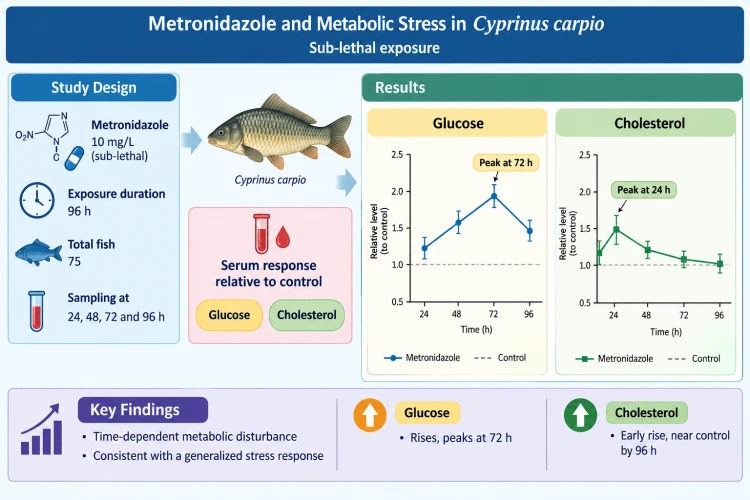
The present study evaluated the temporal effects of metronidazole exposure on plasma glucose and cholesterol levels in common carp (Cyprinus carpio). A total of 75 fish were allocated to independent experimental cohorts (one control group and four exposure groups) to evaluate their biochemical responses at 24, 48, 72, and 96 h postexposure to a sublethal metronidazole concentration of 10 mg/L. Compared with those in the control group, the plasma glucose concentrations in the treatment group increased significantly at 24 and 48 h, reaching a maximum hyperglycemic peak at 72 h. Although a decrease in glucose was observed at 96 h, the values remained significantly elevated compared with those in the control group, suggesting a sustained stress response. Conversely, plasma cholesterol exhibited a distinct, contrasting temporal profile; a marked, significant increase occurred at 24 h postexposure, followed by a progressive, stepwise decrease at 48 and 72 h, ultimately returning to near-control baseline values by 96 h. The early spike in cholesterol is likely attributable to the stress-induced mobilization of hepatic lipid reserves, while the subsequent normalization suggests the activation of compensatory regulatory mechanisms. Collectively, these findings indicate that sublethal metronidazole exposure induces acute, time-dependent perturbations in the carbohydrate and lipid metabolism of C. carpio, which is consistent with a generalized physiological stress response, highlighting the clear adaptive capacity of the species during prolonged exposure.
Keywords
Graphical Abstract
Novelty Statement
This study provides the high-resolution temporal profile of sublethal metronidazole exposure on carbohydrate and lipid metabolism in Cyprinus carpio. By tracking independent cohorts over 96 hours, we reveal distinct, contrasting metabolic responses: sustained hyperglycemia peaking at 72 hours versus a rapid, transient spike in plasma cholesterol that normalizes via compensatory mechanisms. These findings highlight the species' specific physiological stress thresholds and adaptive capacity during acute pharmaceutical exposure.
1. Introduction
Antibiotics are used to treat bacterial infections in human and veterinary medicines and are also utilized as feed supplements in industrial livestock production, causing fears regarding their impact on the environment, especially on the aquatic environment.[1] Approximately 1.8 lakh tones of antibiotics were consumed globally in 2020, with China accounting for more than 55%, and 1.31 lakh tonnes used in food animals, projected to increase to 2.37 lakh tones by 2030.[2] In high-income countries, the United States of America, France, and Italy are major antibiotic consumers, whereas China, India, and Pakistan lead in low- and middle-income countries, with approximately 300 million people aged 15–64 years using antibiotics annually before COVID-19.[3] The word “antibiotics” comes from two parts: “anti”, meaning against, and “biosis”, meaning “life”, referring to substances that act against microbes. Previously, antibiotics were defined as substances produced by one microorganism to inhibit another, but they now include both natural and synthetic compounds.[4] Hundreds of antibiotics and antimycotic substances are used in human and veterinary medicine, with more than 250 in Germany alone.[5] Uniform international data on antibiotic use are lacking, and the existing information is heterogeneous, reflecting different usage patterns worldwide.[6] Antibiotic use, whether prescribed or not, varies widely among different nations.[7] Even within a single country such as Germany, antibiotic consumption varies regionally, ranging from 9.6 to 17.3 DDDs per capita per day.[8] The antibiotics permitted for use in aquaculture include oxytetracycline, florfenicol, erythromycin, sarafloxacin, premixes, and sulfonamides enhanced with trimethoprim or orrimethoprim.[9] A study by Grave et al. (2008) examined the use of antimicrobial drugs in Norwegian aquaculture between 2000 and 2005.[10] The synthetic broad-spectrum antimicrobial metronidazole is widely utilized and frequently administered in veterinary medicine to treat animals of all sizes.[11] It is effective against anaerobic bacteria and protozoa responsible for diseases such as giardiasis, amoebiasis, bowel infections, and peritonitis and is widely used as a postoperative prophylactic treatment.[12] In the liver, metronidazole is metabolized by oxidation and glucuronidation and is eliminated mainly through renal excretion.[13] Metronidazole can induce neurological side effects, particularly when it is used at high doses or for extended durations.[14]
Biochemical analysis serves as an essential method for determining the physiological and metabolic responses of organisms under both standard and stress conditions.[15] It involves the comprehensive quantification of biomolecules, including carbohydrates, proteins, lipids, and enzymes, that are indispensable for cellular integrity, organization, and physiological activity.[16] Fluctuations, alterations, and variations in biochemical parameters are recognized as valuable biomarkers for detecting environmental stress, toxicity, and disease-related conditions.[17] The escalation and accumulation of environmental pollution in recent decades, particularly from pharmaceutical residues, have drawn considerable attention because of their disruptive effects on biochemical and physiological processes in living systems.[18] The tendency of aquatic ecosystems to act as final sinks makes them highly vulnerable to the accumulation of diverse anthropogenic pollutants.[19] Biochemical markers such as glucose and cholesterol are typically employed to assess metabolic disturbances induced by toxic substances.[20] The occurrence of increased glucose levels during stress conditions is commonly linked to hyperglycemia, which is regulated by endocrine responses, particularly cortisol secretion.[21] Similarly, fluctuations and changes in cholesterol levels serve as important markers of disrupted lipid metabolism and impaired membrane integrity in response to toxic conditions.[22] As a result, biochemical analysis functions as a reliable, efficient, and effective technique for determining the toxicological effects of environmental contaminants.[23] Fish are particularly vulnerable to antibiotic contamination because of their continuous exposure to waterborne pollutants, making them valuable bioindicators for ecotoxicological assessment. Among freshwater species, the common carp (Cyprinus carpio) is extensively used as a model organism owing to its ecological relevance, wide distribution, and sensitivity to environmental stressors.[24,25] Cyprinus carpio belongs to the order Cypriniformes and the family Cyprinidae - the largest family of freshwater fishes and inhabits diverse freshwater ecosystems, such as ponds, lakes, and rivers, with occasional occurrence in brackish environments.[26] Its broad geographical distribution, particularly across Asia and parts of Europe, further supports its suitability as a model species in aquatic toxicology studies.[27-29] Earlier work on metronidazole in common carp has mainly addressed hepatic oxidative stress and tissue damage. This study instead follows serum glucose and cholesterol at four consecutive time points which shows how the response develops and partly recovers rather than giving a single end-point value. This study focuses on evaluating and investigating selected biochemical parameters to determine their role as potential and reliable biomarkers of toxic stress in aquatic organisms.[30]
2. Materials and methods
The common carp (Cyprinus carpio) used in this study was obtained from the fish farm of the Department of Fisheries, CSK Himachal Pradesh Agricultural University, Palampur, India. The experimental animals had an average length of 18.4 ± 4.1 cm and an average weight of 88 ± 10 g. Prior to the experimental trials, the fish were preventively treated with a 0.2% potassium permanganate (KMnO4) solution for 2 minutes to eliminate potential dermal ectoparasites. A total of 75 fish were transferred to the laboratory environment and acclimatized for 15 days in glass aquaria containing 50 L of dechlorinated water. Throughout the acclimation and experimental periods, the water temperature was maintained at 18–20°C, and the pH ranged between 7.6 and 7.8. Dissolved oxygen was supplied continuously via mechanical aeration pumps to maintain levels consistently above 5.0 mg/L, preventing hypoxic stress. The aquaria were kept under a natural seasonal photoperiod (approximately 12 h light:12 h dark cycle). The fish were fed a farm-processed pelleted diet once daily, and 100% water exchange was performed every 24 hours to eliminate metabolic excretions. The experimental architecture comprised five distinct allocations of 15 fish each (each allocation consisting of 3 replicate aquaria with 5 fish per replicate): one designated control group and four independent metronidazole exposure groups corresponding to the specific sampling intervals (24, 48, 72, and 96 hours). For the exposure cohorts, metronidazole (Sachin Parenteral Pvt. Ltd., liquid formulation, 500 mg/100 mL) was administered at a sublethal concentration of 10 mg/L, which was determined on the basis of established protocols by Çavaş and Ergene‐Gözükara (2005).[31] The four exposure groups were subjected to the drug for their respective durations (24, 48, 72, or 96 h) prior to terminal sampling. To evaluate the chronological biochemical response without the confounding effects of repetitive handling stress, sampling was executed strictly without replacement. At each designated time point (24, 48, 72, and 96 h), exactly 3 fish were randomly selected from the active exposure group (1 fish per replicate aquarium (n = 3)). Simultaneously, 3 fish were sampled from the single shared control group (1 fish per replicate aquarium (n = 3)). Consequently, out of the 15 total fish in the control group, 12 unique individuals were utilized across the four time points, while the remaining 3 fish served as unsampled environmental reserve animals. No individual fish was subjected to repeated blood extraction. Animal handling, experimental design, and data reporting in this study adhered to the ARRIVE guidelines to ensure transparency and reproducibility.
2.1 Protocol for biochemical analysis
Blood samples were collected from the caudal vein of uneuthanized fish using heparinized syringes. To isolate plasma, blood samples were immediately centrifuged at 10,000 rpm for 10–15 min using a Tarson Spinning MC-00 centrifuge at room temperature. Plasma glucose and cholesterol levels were quantified using commercial diagnostic assay kits (Agappe Diagnostics Ltd., India) calibrated for serum/plasma matrices and read on a Vchem Next Biochemistry Analyzer in strict accordance with the manufacturer's operational protocols.
2.2 Statistical analysis
All data points are expressed as the mean ± standard error (SE). Variations in plasma glucose and cholesterol across the groups and intervals were evaluated using one-way analysis of variance (ANOVA), followed by Tukey’s honestly significant difference (HSD) post hoc test for multiple comparisons. Statistical computations were performed using Minitab software, with the threshold for statistical significance set a priori at p < 0.05.
3. Results
Compared with the control group, exposure to 10 mg/L metronidazole resulted in significant and time-dependent alterations in the plasma biochemical parameters of Cyprinus carpio (p < 0.05). The plasma glucose levels tended to increase steadily and significantly during the initial phases of exposure. A significant increase was observed as early as 24 h postexposure compared with the control baseline, followed by a sharp and distinct increase at 48 h. The maximum hyperglycemic response was recorded at 72 h (p < 0.05). Although the glucose levels subsequently decreased at the 96 h interval, they remained significantly higher than those of the control group did (Fig. 1). This overall kinetic profile indicates a robust hyperglycemic response triggered by acute physiological stress during sustained chemical exposure.
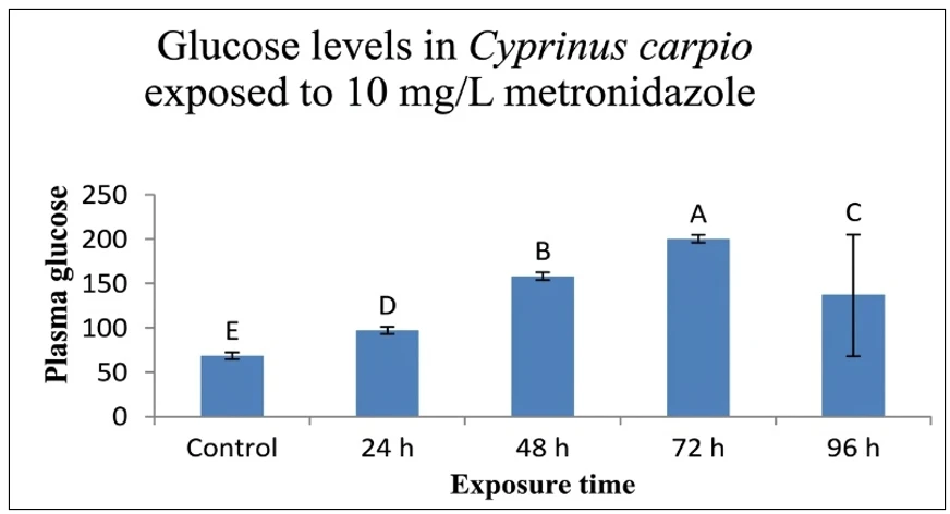
Fig. 1: Effect of 10 mg/L metronidazole exposure on plasma glucose levels in Cyprinus carpio at different time intervals. Data points represent the mean ± standard error (n = 3). Columns sharing distinct superscript letters indicate statistically significant differences (p < 0.05) according to one-way ANOVA followed by Tukey's HSD post hoc test.
Plasma cholesterol levels demonstrated a contrasting temporal pattern of metabolic disruption. A sharp, rapid increase was detected at 24 h, with the highest overall value observed across all the exposure windows (p < 0.05). This initial spike was followed by a progressive, stepwise decline at 48 h and 72 h; however, concentrations during these intervals remained significantly elevated relative to those in the control cohort. By the conclusion of the exposure trial at 96 h, the plasma cholesterol levels had returned to baseline values, and the difference from the control group was not statistically significant (Fig. 2). This trend highlights an acute initial metabolic disturbance followed by a clear phase of partial physiological recovery.
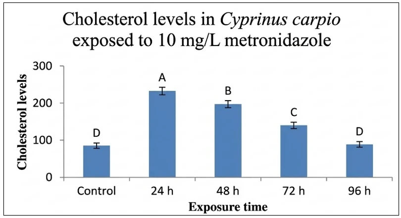
Fig. 2: Effect of 10 mg/L metronidazole exposure on plasma cholesterol levels in Cyprinus carpio at different
time intervals. Data points represent the mean ± standard error (n = 3). Columns sharing distinct superscript
letters indicate statistically significant differences (p < 0.05) according to one-way ANOVA followed by Tukey's HSD post hoc test.
4. Discussion
These findings confirm that sublethal exposure to metronidazole (10 mg/L) significantly alters glucose and cholesterol levels in a time-dependent manner in Cyprinus carpio. These alterations and variations indicate metabolic stress and adaptive, compensatory physiological responses to pharmaceutical exposure, highlighting the effectiveness of biochemical indices as reliable biomarkers in aquatic toxicology. Metronidazole toxicity is closely associated with its intracellular reductive activity, where enzymatic nitroreduction produces nitro-radical anions and other reactive intermediates.[32] The generated reactive metabolites can bind to and damage DNA or undergo redox cycling, thereby promoting the excessive formation of ROS such as superoxide radicals and hydrogen peroxide. Metronidazole increases the levels of malondialdehyde, glutathione, superoxide dismutase and catalase in the liver of common carp and induces hepatic oxidative stress and tissue damage.[33] In another study on carp, metronidazole was found to cause moderate vacuolation, cholestasis, cytoplasmic degeneration and hepatocellular necrosis.[34] The main reason for metronidazole toxicity is that it involves reductive activation, which alters the state, carbon flow and ribonucleotide reductase activity and provides a mechanistic basis for metabolic disruption.[35] The gradual increase in glucose levels over time, along with a continuous increase during prolonged exposure, suggests a stress-induced hyperglycemic response. Toxicant exposure in fish often leads to increased glucose levels as a result of HPI axis activation and elevated cortisol and catecholamine secretion.[36] The stimulation of glycogenolysis and gluconeogenesis by these hormones contributes to increased blood glucose concentrations.[37] Studies have reported similar increases in glucose levels in Cyprinus carpio subjected to pesticides, heavy metals, and pharmaceutical substances.[38,39] The highest glucose level at 72 h indicates a pronounced stress response, which is likely associated with enhanced mobilization and use of energy reserves to counter toxic stress.[40] Even though the levels were still significantly greater than those in the control, the observed decline at 96 h may suggest physiological adaptation or depletion of stored energy.[41] Fish subjected to prolonged toxicant exposure often display biphasic or two-phase physiological responses.[17] When fish are exposed to antibiotics, glycogen breakdown is triggered in their liver, suggesting that carbohydrate consumption meets energy demand. In a study on zebrafish, they were exposed to a rubber-derived toxicant called 6PPD-quinone. The levels of glucose and glycogen in the liver increase, whereas the levels of pyruvic acid decrease, suggesting the inhibition of glycolysis and a shift toward glycogen synthesis.[42] The expression of genes essential for glucose metabolism and insulin secretion, such as those encoding hexokinase and glucokinase, is suppressed, indicating that chemically induced metabolic disturbance and impaired insulin signaling and glucose–lipid balance. Cholesterol levels increased significantly at 24 h but gradually decreased to control levels by 96 h, indicating its involvement in membrane stability, steroidogenesis, and lipid metabolism.[43] The increase in cholesterol levels in fish under toxicant stress suggests alterations in lipid metabolism and hepatic impairment.[44] The gradual decrease in cholesterol levels during extended exposure may suggest metabolic adaptation and the use of lipids as alternative energy sources under stress.[45] The restoration of cholesterol levels toward near-control values at 96 h may indicate the activation of compensatory mechanisms and partial recovery of metabolic homeostasis.[17] These recovery trends are frequently reported in fish exposed to sublethal doses of xenobiotics, whose recovery patterns are similar under such stress conditions.[38] An environmentally detected pharmaceutical substance, metformin, disrupted the mitochondrial electron transport chain and tricarboxylic acid cycle in zebrafish, directly affecting energy metabolism. Metformin exposure disrupts cholesterol and fatty acid biosynthesis, altering the hepatosomatic index and expression of lipid metabolism genes such as hmgcra.[46] Similarly, the triazole fungicide triflumizole upregulated the expression of genes related to lipid synthesis in zebrafish.[47] In a study on rats, metronidazole was found to affect genes that help the liver regulate cholesterol and triglycerides.[48] The observed biochemical variations may be indicative of oxidative stress, reflecting its role in the study outcomes. Like other pharmaceutical contaminants, metronidazole has been shown to stimulate the generation of reactive oxygen species (ROS), resulting in oxidative damage in aquatic organisms.[49] Disruptions in cellular metabolism, enzymatic function, and membrane stability caused by oxidative stress can significantly influence biochemical parameters.[50] The hyperglycemia and changes in lipid levels observed in this study may be linked to liver dysfunction caused by metronidazole exposure. As a crucial organ in carbohydrate and lipid metabolism, the liver can markedly alter its biochemical parameters when it is affected by toxicant-induced damage.[51] Studies in fish have demonstrated that exposure to antibiotics and pharmaceutical compounds can impair liver function, leading to alterations in glucose and cholesterol levels.[4]
5. Conclusion
Acute exposure of Cyprinus carpio to metronidazole at 10 mg/L produced clear, time-dependent changes in
serum glucose and cholesterol over 96 h. Glucose rose progressively, peaked at 72 h, and declined slightly at 96 h while remaining significantly above control. This is consistent with a stress-induced hyperglycaemic response. Cholesterol showed a different pattern: it peaked at 24 h, fell gradually at 48 and 72 h, and returned to near-control values by 96 h, suggesting an early disturbance of lipid metabolism followed by partial compensation. Together, these results show that glucose and cholesterol respond sensitively to metronidazole and can serve as early, low-cost indicators of metabolic stress in carp. The mechanisms proposed here, including oxidative stress and hepatic involvement, are inferred from earlier studies and need to be confirmed with enzymatic, oxidative-stress and histopathological endpoints, and across a range of concentrations.
Acknowledgment
The authors gratefully acknowledge the State Department of Fisheries, Himachal Pradesh (https://himachal.nic.in/fisheries) to provide the financial support necessary to carry out the present research work. The funding provided by the Department is sincerely appreciated, as it facilitated the successful completion of this study.
Funding Declaration
The study received financial support from the State Department of Fisheries, Himachal Pradesh (State Adhoc Misc. 2348-46).
Institutional Review Board Statement
This study was conducted as part of a formal research project sanctioned and funded by the State Department of Fisheries Himachal Pradesh (State Adhoc Misc. 2348-46) and administratively approved by Chaudhary Sarwan Kumar Himachal Pradesh Krishi Vishvavidyalaya. Because this project was processed as an official state agricultural/fisheries monitoring initiative, an individual, prospective protocol number from the Institutional Animal Ethics Committee (IAEC) was not obtained prior to execution. However, all experimental procedures, including 96-hour metronidazole exposure, preventive KMnO₄ treatments, and serial blood sampling, were conducted within university facilities in strict accordance with the baseline institutional animal welfare standards and national guidelines prescribed by the Committee for the Control and Supervision of Experiments on Animals (CCSEA), India. The authors confirm that all efforts were made to minimize animal suffering and optimize housing conditions throughout the trial.
Data Availability Statement
The datasets generated and/or analyzed during the current study that support the findings are available from the corresponding author upon reasonable request.
Conflict of Interest
There is no conflict of interest.
Artificial Intelligence (AI) Use Disclosure
The authors declare that artificial intelligence (AI)-assisted tools were used only for language refinement, grammar improvement, and manuscript structuring purposes during the preparation of this work. All technical content, experimental implementation, results, and interpretations were independently developed and verified by the authors.
Supporting Information
Not applicable.
References
- [1] M. Gonzalez-Pleiter, S. Gonzalo, I. Rodea-Palomares, F. Leganes, R. Rosal, K. Boltes, E. Marco, F. Fernandez-Pinas, Toxicity of five antibiotics and their mixtures towards photosynthetic aquatic organisms: implications for environmental risk assessment, Water Research, 2013, 47, 2050-2064, doi: 10.1016/j.watres.2013.01.020.
- [2] R. Mulchandani, Y. Wang, M. Gilbert, T. P. Van Boeckel, Global trends in antimicrobial use in food-producing animals: 2020 to 2030, PLOS Global Public Health, 2023, 3, e0001305, doi: 10.1371/journal.pgph.0001305.
- [3] O. P. Bansal, Antibiotics in the environment: A review, International Journal of Frontiers in Biology and Pharmacy Research, 2022, 2, 057–083, doi: 10.53294/ijfbpr.2022.2.1.0030.
- [4] K. Kümmerer, The presence of pharmaceuticals in the environment due to human use–present knowledge and future challenges, Journal of Environmental Management, 2009, 90, 2354-2366, doi: 10.1016/j.jenvman.2009.01.023.
- [5] K. Kümmerer, A. Henninger, Promoting resistance by the emission of antibiotics from hospitals and households into effluent, Clinical Microbiology and Infection, 2003, 9, 1203-1214, doi: 10.1111/j.1469-0691.2003.00739.x.
- [6] K. Kümmerer, Pharmaceuticals in the environment–a brief summary. In Pharmaceuticals in the environment: Sources, fate, effects and risks, Berlin, Heidelberg: Springer Berlin Heidelberg, 2008, 3-21.
- [7] S. L. Bronzwaer, O. Cars, U. Buchholz, S. Mölstad, W. Goettsch, I. K. Veldhuijzen, J. L. Kool, M. J. Sprenger, J. E. Degener, The relationship between antimicrobial use and antimicrobial resistance in Europe, Emerging Infectious Diseases, 2002, 8, 278, doi: 10.3201/eid0803.010192.
- [8] K. De With, H. Schroder, E. Meyer, K. Nink, S. Hoffmann, M. Steib-Bauert, R. Kammerer, S. Ruess, F. D. Daschner, W. V. Kern, Antibiotic use in Germany and Europe, Deutsche Medizinische Wochenschrift, 2004, 129, 1987-1992, doi: 10.1055/s-2004-831838.
- [9] P. H. Serrano, Responsible Use of Antibiotics in Aquaculture, FAO Fisheries Technical Paper No. 469, Food and Agriculture Organization of the United Nations, Rome, 2005.
- [10] K. Grave, M. K. Hansen, H. Kruse, M. Bangen, A. B. Kristoffersen, Prescription of antimicrobial drugs in Norwegian aquaculture with an emphasis on “new” fish species, Preventive Veterinary Medicine, 2008, 83, 156-169, doi: 10.1016/j.prevetmed.2007.07.002.
- [11] G. P. Mark, Veterinary Drugs, 4th ed., Elsevier, New York, 2016, pp. 524–527.
- [12] D. C. Plumb, Plumb’s Veterinary Drug Handbook, 6th ed., Blackwell Publishing, Iowa, 2008, 2610–2613.
- [13] D. Leitsch, A review on metronidazole: An old warhorse in antimicrobial chemotherapy, Parasitology, 2019, 146, 1167–1178, doi: 10.1017/S0031182017002025.
- [14] L. Li, X. Tang, W. Li, S. Liang, Q. Zhu, M. Wu, A case of methylprednisolone treatment for metronidazole-induced encephalopathy, BMC Neurology, 2019, 19, 49, doi: 10.1186/s12883-019-1278-6.
- [15] D. L. Nelson, M. M. Cox, Lehninger Principles of Biochemistry, 7th ed., W. H. Freeman, 2017.
- [16] J. M. Berg, J. L. Tymoczko, G. J. Gatto Jr, L. Stryer, Biochemistry, 8th Edition, W.H. Freeman & Company, New York, 2015.
- [17] R. van der Oost, J. Beyer, N. P. E. Vermeulen, Fish bioaccumulation and biomarkers in environmental risk assessment: A review, Environmental Toxicology and Pharmacology, 2003, 13, 57–149, doi: 10.1016/S1382-6689(02)00126-6.
- [18] K. Fent, A. A. Weston, D. Caminada, Ecotoxicology of human pharmaceuticals, Aquatic Toxicology, 2006, 76, 122-159, doi: 10.1016/j.aquatox.2005.09.009.
- [19] K. Kümmerer, Antibiotics in the aquatic environment–a review–part I, Chemosphere, 2009, 75, 417-434, doi: 10.1016/j.chemosphere.2008.11.086.
- [20] J. V. Rao, Biochemical alterations in euryhaline fish, Oreochromis mossambicus, exposed to sub-lethal concentrations of an organophosphorus insecticide, monocrotophos, Chemosphere, 2006, 65, 1814–1820, doi: 10.1016/j.chemosphere.2006.04.015.
- [21] G. A. Wedemeyer, B. A. Barton, D. J. McLeay, Stress and acclimation, in Methods for Fish Biology, American Fisheries Society, Bethesda, Maryland, 1990, 451–489.
- [22] S. S. Vutukuru, Acute effects of hexavalent chromium on survival, oxygen consumption, hematological parameters and some biochemical profiles of the Indian major carp, Labeo rohita, International Journal of Environmental Research and Public Health, 2005, 2, 456–462, doi: 10.3390/ijerph2005030010.
- [23] C. H. Walker, S. P. Hopkin, R. M. Sibly, D. B. Peakall, Principles of Ecotoxicology, 4th ed., CRC Press, 2012.
- [24] M. Hejna, D. Kapuścińska, A. Aksmann, Pharmaceuticals in the aquatic environment: A review on eco-toxicology and the remediation potential of algae, International Journal of Environmental Research and Public Health, 2022, 19, 7717, doi: 10.3390/ijerph19137717.
- [25] A. Sharma, M. Sharma, T. Shah, P. Dhar, Effect of metronidazole on hematological parameters in common carp (Cyprinus carpio), Pantnagar Journal of Research, 2024, 22, 443–448.
- [26] V. Barus, Cyprinus carpio (Linnaeus 1758), The freshwater fishes of Europe, 200, 5, 85-179.
- [27] J. Kloskowski, Impact of common carp Cyprinus carpio on aquatic communities: direct trophic effects versus habitat deterioration, Fundamental and Applied Limnology-Archiv furHydrobiologie, 2011, 178, 245, doi: 10.1127/1863-9135/2011/0178-0245.
- [28] J. I. Parkos, D. Wahl, Effects of common carp (Cyprinus carpio), an exotic fish, on aquatic ecosystems. Ilinois Natural History Survey report of January/February 2000. University of Illinois Board of Trustees. Center for Aquatic Ecology, 2014.
- [29] M. J. Weber, M. L. Brown, Relationships among invasive common carp, native fishes and physicochemical characteristics in upper Midwest (USA) lakes, Ecology of Freshwater Fish, 2011, 20, 270–278, doi: 10.1111/j.1600-0633.2011.00493.x.
- [30] R. K. Trivedi, P. K. Goel, C. L. Trisal, Practical Methods in Ecology and Environmental Science, 2009.
- [31] T. Çavaş, S. Ergene‐Gözükara, Micronucleus test in fish cells: a bioassay for in situ monitoring of genotoxic pollution in the marine environment, Environmental and Molecular Mutagenesis, 2005, 46, 64-70, doi: 10.1002/em.20130.
- [32] D. I. Edwards, Nitroimidazole drugs-action and resistance mechanisms I. Mechanism of action, Journal of Antimicrobial Chemotherapy, 1993, 31, 9-20, doi: 10.1093/jac/31.1.9.
- [33] J. Han, H. Cai, J. Wang, G. Liu, Detrimental effects of metronidazole on the liver of freshwater common carp (Cyprinus carpio L.), Bulletin of Environmental Contamination and Toxicology, 2013, 91, 444-449, doi: 10.1007/s00128-013-1059-7.
- [34] M. Waqar, U. Elahi, S. Masud, S. Shahzadi, S. Yousaf, and A. A. Hasaan, Effect of Antibiotics Exposure on Gills, Liver, Kidney and Brain of Common Carp (Cyprinus carpio), Indus Journal of Bioscience Research, 2025, 3, 263–273, doi: 10.70749/ijbr.v3i5.1232.
- [35] S. A. Dingsdag, N, Metronidazole: an update on metabolism, structure-cytotoxicity and resistance mechanisms, Journal of Antimicrobial Chemotherapy, 2018, 73, 265–279, doi: 10.1093/jac/dkx351.
- [36] B. A. Barton, Stress in fishes: a diversity of responses with particular reference to changes in circulating corticosteroids, Integrative and Comparative Biology, 2002, 42, 517-525, doi: 10.1093/icb/42.3.517.
- [37] T. P. Mommsen, M. M. Vijayan, T. W. Moon, Cortisol in teleosts: Dynamics, mechanisms of action, and metabolic regulation, Reviews in Fish Biology and Fisheries, 1999, 9, 211–268, doi: 10.1023/A:1008924418720.
- [38] M. Banaee, A. Sureda, A. R. Mirvaghefi, K. Ahmadi, Effects of diazinon on biochemical parameters of blood in rainbow trout (Oncorhynchus mykiss), Pesticide Biochemistry and Physiology, 2011, 99, 1-6, doi: 10.1016/j.pestbp.2010.09.001.
- [39] M. Sevcikova, H. Modrá, A. Slaninova, Z. Svobodova, Metals as a cause of oxidative stress in fish: a review, Veterinární Medicína, 2011, 56, 537-546.
- [40] S. E. Wendelaar Bonga, The stress response in fish, Physiological Reviews, 1997, 77, 591–625, doi: 10.1152/physrev.1997.77.3.591.
- [41] A. K. Nayak, Impact of environmental stress on fish, World Journal of Fish and Marine Sciences, 2010, 2, 451–454.
- [42] F. Jiao, Y. Zhao, Q. Yue, Q. Wang, Z. Li, W. Lin, L. Han, L. Wei, Chronic toxicity mechanisms of 6PPD and 6PPD-Quinone in zebrafish, Environmental Science and Ecotechnology, 2025, 25, 100567, doi: 10.1016/j.ese.2025.100567.
- [43] N. W. Tietz, Clinical Guide to Laboratory Tests, Saunders, 1995.
- [44] M. Abdel‐Tawwab, H. S. Hamed, Effect of bisphenol A toxicity on growth performance, biochemical variables, and oxidative stress biomarkers of Nile tilapia, Oreochromis niloticus (L.), Journal of Applied Ichthyology, 2018, 34, 1117-1125, doi: 10.1111/jai.13763.
- [45] O. K. Adeyemo, Haematological and histopathological effects of cassava mill effluent in Clarias gariepinus, African Journal of Biomedical Research, 2005, 8, 179-183.
- [46] S. Barros, M. Ribeiro, A. M. Coimbra, M. Pinheiro, H. Morais, N. Alves, R. Montes, R. Rodil, J. B. Quintana, M. M. Santos, T. Neuparth, Metformin disrupts Danio rerio metabolism at environmentally relevant concentrations: A full life-cycle study, Science of The Total Environment, 2022, 846, 157361, doi: 10.1016/j.scitotenv.2022.157361.
- [47] L. Bai, P. Shi, K. Jia, H. Yin, J. Xu, X. Yan, K. Liao, Triflumizole induces developmental toxicity, liver damage, oxidative stress, heat shock response, inflammation, and lipid synthesis in zebrafish, Toxics, 2022, 10, 698, doi: 10.3390/toxics10110698.
- [48] A. Eguchi, S. Mizukami, M. Nakamura, S. Masuda, H. Murayama, M. Kawashima, M. Inohana, R. Nagahara, M. Kobayashi, R. Yamashita, S. Uomoto, Metronidazole enhances steatosis-related early-stage hepatocarcinogenesis in high fat diet-fed rats through DNA double-strand breaks and modulation of autophagy, Environmental Science and Pollution Research, 2022, 29, 779-789, doi: 10.1007/s11356-021-15689-2.
- [49] L. Grădinariu, M. Crețu, C. Vizireanu, L. Dediu, Oxidative stress biomarkers in fish exposed to environmental concentrations of pharmaceutical pollutants: A review, Biology, 2025, 14, 472, doi: 10.3390/biology14050472.
- [50] D. R. Livingstone, Contaminant-stimulated reactive oxygen species production and oxidative damage in aquatic organisms, Marine Pollution Bulletin, 2001, 42, 656–666, doi: 10.1016/S0025-326X(01)00060-1.
- [51] D. Schlenk, J. Goldstone, M. O. James, P. van den Hurk, Biotransformation in fishes. In Toxicology of fishes, CRC Press, 2024, 60-120.
Publisher Note
The views, statements, and data in all publications solely belong to the author. GR Scholastic is not responsible for any injury resulting from the ideas, methods, or products mentioned. GR Scholastic remains neutral regarding jurisdictional claims in published maps and institutional affiliations.
Open Access
This article is licensed under a Creative Commons Attribution-Non Commercial 4.0 International License, which permits the non-commercial use, sharing, adaptation, distribution and reproduction in any medium or format, as long as appropriate credit to the original author(s) and the source is given by providing a link to the Creative Commons License and changes need to be indicated if there are any. The images or other third-party material in this article are included in the article's Creative Commons License, unless indicated otherwise in a credit line to the material. If material is not included in the article's Creative Commons License and your intended use is not permitted by statutory regulation or exceeds the permitted use, you will need to obtain permission directly from the copyright holder. To view a copy of this License, visit: https://creativecommons.org/licenses/by-nc/4.0/
© The Author(s) 2026