

 Open Access

Abstract
Esophageal foreign bodies are potentially life-threatening emergencies in cats that require prompt diagnosis and intervention. This report describes the successful endoscopic removal of a fishhook from the thoracic esophagus of a 4-year-old intact male domestic cat weighing 3.9 kg, which was presented to Paws N Claws Veterinary Clinic, Brunei Darussalam, after which accidental ingestion occurred. The cat exhibited mild hypersalivation, and thoracic radiography confirmed that a radiopaque fishing hook was lodged in the thoracic esophagus. Under general anesthesia induced with tiletamine–zolazepam and maintained with sevoflurane in oxygen, endoscopy revealed that the hook was mildly attached to the esophageal wall upon insufflation of the air. Using foreign-body retrieval forceps through the 2.0-mm working channel, controlled gentle countertraction was used to successfully disengage and retrieve the hook without further manipulation. The esophageal mucosa was subsequently reexamined endoscopically for residual foreign material, and the absence of a tear was confirmed. Postoperatively, the cat received supportive care, including anti-inflammatory and antiemetic medications, alongside close clinical monitoring. The cat recovered uneventfully, with normal appetite, swallowing function, and physical activity at the three-day follow-up examination. This case demonstrates that endoscopic retrieval can provide a safe, minimally invasive, and effective approach for the removal of esophageal fishing hooks in cats when performed with careful visualization and controlled traction.
Keywords
Graphical Abstract
1. Introduction
Esophageal foreign bodies are important gastrointestinal emergencies frequently encountered in feline and canine practice and can result in significant morbidity if not diagnosed and treated promptly. Compared with cats, esophageal foreign bodies are more frequently encountered in dogs.[1] Common foreign objects found within the esophagus and gastrointestinal tract of small animals include bones and cartilage fragments, fishhooks, balls, toys, needles, elastic hair bands, and trichobezoars.[2] The diagnosis of an esophageal foreign body is established through a combination of clinical findings, patient history, and diagnostic imaging. Plain radiography may help identify radiopaque objects, whereas endoscopy allows direct visualization of the esophageal lumen, mucosa, and retained foreign material.[3] Clinical manifestations depend on the location, size, shape, and duration of foreign body impact. Affected animals may exhibit various clinical signs, including excessive salivation, repeated gagging or retching, vomiting, regurgitation, reduced appetite, signs of discomfort or pain, and varying degrees of respiratory difficulty.[4] The success rate of endoscopic foreign body retrieval varies depending on the location of the foreign body, with successful removal reported in approximately 68–88% of esophageal cases and 78–94% of gastric cases.[5] When endoscopic retrieval of an esophageal foreign body is unsuccessful, surgical intervention may be necessary. However, in selected cases involving digestible materials, the foreign body may be gently advanced into the stomach rather than extracted directly. Esophageal foreign bodies requiring surgical intervention have been associated with prolonged hospitalization and an increased risk of mortality.[5] Given its minimally invasive nature and therapeutic advantages, endoscopy represents a valuable alternative to surgical intervention for the management of suitable esophageal foreign bodies. Therefore, this case report describes the successful endoscopic retrieval of an esophageal foreign body in a cat, highlighting the clinical approach, endoscopic procedure, and treatment outcome.
2. Case presentation
A 4-year-old intact male nondescript cat weighing 3.9 kg was presented to Paws N Claws Veterinary Care,
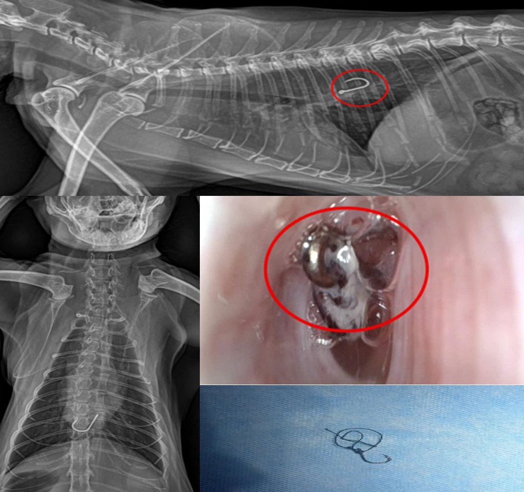
Kuilap, Brunei Darussalam, following an episode of accidental ingestion of a fishing hook, which was witnessed by the owner. Mild hypersalivation was observed at presentation. On physical examination, the cat was bright and alert, with a rectal temperature of 39.5°C. Plain thoracic radiography was performed to determine the location of the suspected foreign body. Radiographs revealed a radiopaque fishing hook lodged within the thoracic portion of the esophagus (Figs. 1 and 2). Prior to the endoscopic procedure, routine hematological and biochemical investigations were performed. All measured parameters were within the respective reference ranges. General anesthesia was induced using tiletamine–zolazepam (Zoletil-100; Virbac AH, Inc.) at 10 mg/kg body weight intramuscularly, after which the cat was positioned for endoscopic examination and retrieval of the esophageal foreign body. The airway was secured using an endotracheal tube, and anesthesia was maintained with sevoflurane (Sevitrue, Abott Healthcare) in oxygen. The vaporizer concentration was adjusted between approximately 2–3% according to the depth of anesthesia and the patient’s physiological parameters. Following the induction of general anesthesia, a portable gastroscope (YH-DWJV007, Yohuvet, China) equipped with a full-HD camera, LED light source, and built-in video recording system was used according to the manufacturer’s instructions. The endoscope had an outer diameter of 5.8 mm, a working length of 750 mm, and a 2.0-mm working channel. The endoscope was introduced gently through the oral cavity and advanced under direct visualization with appropriate air insufflation. It was guided through the pharynx into the cervical and thoracic esophagus.
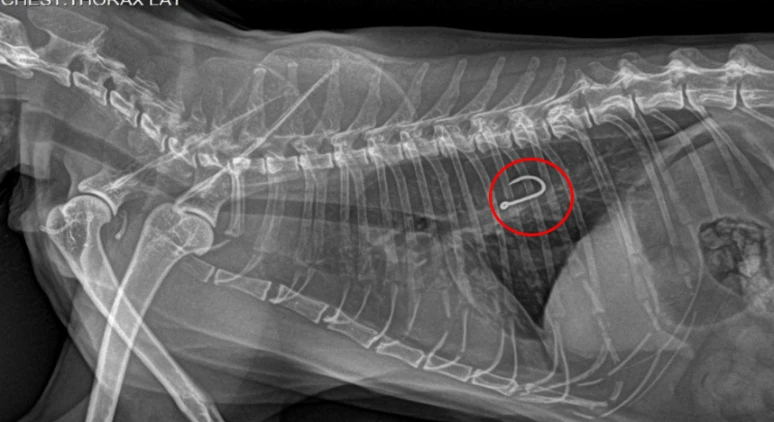
Fig. 1:Left lateral plain radiograph demonstrating a fish hook (circle) lodged within the thoracic esophagus.
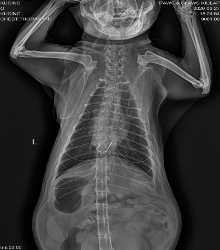
Fig. 2:Ventro-dorsal radiograph demonstrating a fish hook (arrow) lodged within the thoracic esophagus.
The animal was handled humanely throughout the diagnostic and therapeutic procedures. All clinical interventions were performed in accordance with standard veterinary practices, with appropriate measures taken to minimize pain, stress, and suffering. Care was taken to provide appropriate supportive and therapeutic management in view of the cat’s dyspnea. All the procedures were performed under general anesthesia, with the appropriate anesthetic depth maintained throughout the procedure. Postprocedural pain management was provided to ensure adequate pain control, patient comfort, and patient welfare during recovery permission to conduct the study, and data were collected at Paws N Claws Veterinary Care, Kuilap, and Brunei Darussalam prior to data collection. The management also authorized the identification of Paws N Claws Veterinary Care in the research manuscript and any related academic presentations or publications. The report was prepared in accordance with the VCARE guidelines.
3. Results
Endoscopic examination revealed a fishhook tip (Fig. 3) mildly attached to the esophageal wall. The esophageal mucosa showed mild erythematous changes. No significant mucosal injury, erosion, ulceration, active bleeding, or obvious perforation was observed during examination. Foreign-body retrieval forceps were introduced through the 2.0-mm working channel and advanced under direct endoscopic visualization toward the exposed portion of the hook. The hook was securely grasped with the retrieval forceps and locked in position. Gentle countertraction was then applied to disengage the hook from the esophageal wall while simultaneously maintaining the position of the endoscope. Once the hook was released from the esophageal wall, it was carefully withdrawn into the endoscopic field. The endoscope and retrieval forceps were subsequently withdrawn slowly and carefully through the esophagus and oral cavity, with the fish hook securely held by the forceps. Controlled traction was applied under continuous endoscopic visualization to retrieve the foreign body through the esophagus and oral cavity while minimizing additional mucosal trauma. The foreign body was successfully retrieved externally (Fig. 4). Following removal, the esophageal mucosa was reexamined endoscopically to assess residual foreign material and mucosal injury. Following the procedure, the cat recovered from anesthesia. Postoperatively, the cat received supportive care, including 0.5 mg/kg intramuscular prednisolone acetate (MSD Animal Health, USA) and subcutaneous maropitant citrate @1 mg/kg (Vet Maro, Vivaldis Animal Health, India) once daily for two days, with close monitoring for signs of esophageal irritation or injury, including dysphagia, regurgitation, excessive salivation, and discomfort, to ensure adequate hydration. The cat resumed normal appetite and swallowing function, and the owner was advised to monitor for signs of recurrent regurgitation, difficulty swallowing, or other complications. The cat was discharged with oral prednisolone @0.5 mg/kg (Tab Prednidale 5, Dechra, UK) and ondansetron @ 0.5 mg/kg (Syrup Petvomi Fix, Oriheal, India), which was prescribed once daily for three days. A follow-up examination was recommended after three days to reassess swallowing function. The cat had an uneventful recovery, with normal appetite, swallowing function, and physical activity on the review day. Owing to owner constraints, a review endoscopic evaluation was not performed.
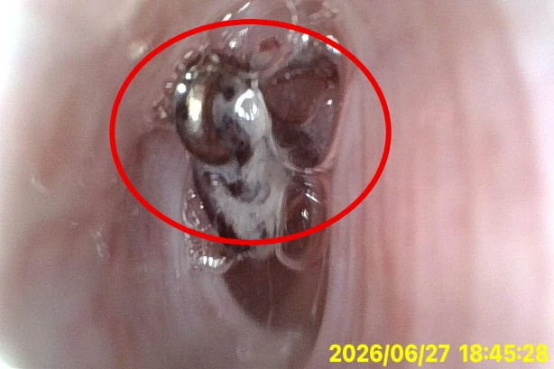
Fig. 3:Endoscopic view of the esophagus showing a fishhook embedded in the esophageal wall.
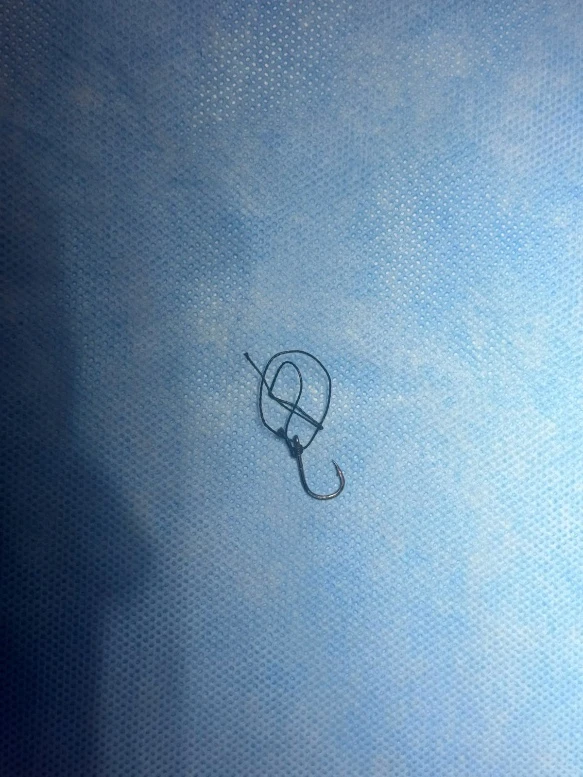
Fig. 4:Fish hook retrieved from the esophagus.
4. Discussion
Gastrointestinal endoscopy enables direct mucosal assessment, biopsy collection, and minimally invasive retrieval of suitable foreign bodies, thereby reducing tissue trauma, postoperative discomfort, and recovery time compared with invasive procedures such as esophagotomy, gastrotomy, or enterotomy. It is particularly valuable in the management of esophageal foreign bodies, which constitute a veterinary emergency because of the prolonged retention, compression, or penetration of the esophageal wall can result in mucosal necrosis, ulceration, and perforation.[4,6] These complications may progress to life-threatening conditions, including pneumothorax, pneumomediastinum, and pyothorax.[7] Therefore, prompt endoscopic diagnosis and removal are essential for minimizing esophageal injury, reducing the need for surgical intervention, and improving patient outcomes. Endoscopy has been widely used worldwide for the retrieval of foreign bodies in cats. In a retrospective study of 52 cats, successful endoscopic removal was achieved in 49 (94.2%) cases, although esophageal foreign bodies were associated with a higher risk of complications.[8] Abd Elkader et al. reported 30 cats with esophageal foreign bodies, including 25 sewing needles and 5 bones; five cases were successfully managed by endoscopy.[8] Similarly, in a series of cats with sewing-needle ingestion, endoscopic removal was successful in 18 of 19 cats with esophageal or gastric needles.[9] However, endoscopy may be hazardous when perforation or severe tissue damage is present; Cariou and Lipscomb reported a cat in which endoscopic retrieval of a perforating esophageal foreign body resulted in pneumomediastinum and pneumothorax, necessitating esophageal resection.[10] Therefore, careful radiographic assessment should be performed before retrieval to determine the location, orientation, and degree of embedding of the foreign body. During endoscopy, the object should be grasped securely and removed with controlled, gentle traction while avoiding excessive manipulation of the esophageal wall. Sharp or deeply embedded foreign bodies warrant particular caution, and the procedure should be discontinued if significant resistance or evidence of perforation is encountered. Immediate surgical intervention is advised in cases of suspected esophageal perforation or unsuccessful endoscopic extraction. To the best of our knowledge, this report represents the first documented case of endoscopic foreign body retrieval in small animal practice in Brunei Darussalam, highlighting its novelty and potential clinical significance.
4. Conclusion
This case demonstrates that endoscopic retrieval can be an effective and minimally invasive technique for the removal of esophageal fishing hooks in cats. Radiographic assessment before endoscopy is essential for determining tumor location and orientation. Careful endoscopic visualization, secure grasping of the foreign body, and controlled, gentle traction are essential to minimize additional trauma to the esophageal mucosa and surrounding tissues. Early diagnosis and intervention, combined with careful postretrieval assessment of the esophageal wall, may further reduce the risk of complications and facilitate an uneventful recovery.
Acknowledgment
The authors would like to thank the staff of Paws N Claws Veterinary Care, Kuilap, Brunei Darussalam, for their assistance in the diagnosis, management, and care of the patient. The authors also sincerely thank the owners for their cooperation throughout the study.
CRediT Author Contribution Statement
Farhan Eravaramkunnath: Conceptualization, Formal analysis, Methodology, Writing – Original draft. Sidharth Singh: Conceptualization, Data Curation. Mangal Singh: Formal analysis, Investigation, Supervision. Anjali Kumari: Writing – Review & editing, Validation, Supervision. Rohin Sharma: Investigation, Supervision. All authors have read and approved the final version of the manuscript for publication and agree to be accountable for all aspects of the work, ensuring that questions related to the accuracy or integrity of any part of the work are appropriately investigated and resolved.
Funding Declaration
This research did not receive any specific grant from funding agencies in the public, commercial, or not-for-profit sectors.
Institutional Review Board Statement
All procedures were conducted in accordance with institutional guidelines and applicable national animal welfare regulations. Given that this was a retrospective case report of routine clinical care, IACUC approval was not needed.
Informed Consent Statement
Informed consent was obtained from the owner for the treatment of the animal.
Consent to publish Statement
Informed consent was obtained from the owner for the publication of the clinical findings and associated information.
Data Availability Statement
The data analyzed during the current study are available from the corresponding author on reasonable request.
Conflict of Interest
There is no conflict of interest.
Artificial Intelligence (AI) Use Disclosure
The authors declare that artificial intelligence (AI)-assisted tools were used only for language refinement, grammar improvement, and manuscript structuring purposes during the preparation of this work. All technical content, experimental implementation, results, and interpretations were independently developed and verified by the authors.
Supporting Information
Not applicable.
References
- [1] P. Gianella, N. S. Pfammatter, I. A. Burgener, Oesophageal and gastric endoscopic foreign body removal: complications and follow‐up of 102 dogs, Journal of Small Animal Practice, 2009, 50, 649-654, doi: 10.1111/j.1748-5827.2009.00845.x.
- [2] G. Maggi, M. Tessadori, M. L. Marenzoni, F. Porciello, D. Caivano, M. C. Marchesi, Endoscopic Retrieval of Esophageal and Gastric Foreign Bodies in Cats and Dogs: A Retrospective Study of 92 Cases. Veterinary Sciences, 2023, 10, 560, doi: 10.3390/vetsci10090560.
- [3] N. A. Abd Elkader, I. A. Emam, H. A. Farghali, M. S. Daghash, N. Y. Salem, Oesophageal foreign bodies in cats: Clinical and anatomic findings, Plos One, 15, 2020, e0233983, doi: 10.1371/journal.pone.0233983.
- [4] B. A. Brisson, S. H. Wainberg, S. Malek, S. Reabel, A. Defarges, W. C. Sears, Risk factors and prognostic indicators for surgical outcome of dogs with esophageal foreign body obstructions, Journal of the American Veterinary Medical Association, 2018, 252, 301-308, doi: 10.2460/javma.252.3.301.
- [5] A. N. Wood, A. E. Gallagher, Survey of instruments and techniques for endoscopic retrieval of esophageal and gastric foreign bodies in cats and dogs, Topics in Companion Animal Medicine, 2021, 45, 100555, doi: 10.1016/j.tcam.2021.100555.
- [6] A. H. Moore, Removal of oesophageal foreign bodies in dogs: use of the fluoroscopic method and outcome, Journal of Small Animal Practice, 2001, 42, 227-230, doi: 10.1111/j.1748-5827.2001.tb02025.x.
- [7] C. Deroy, J. B. Corcuff, F. Billen, A. Hamaide, Removal of oesophageal foreign bodies: comparison between oesophagoscopy and oesophagotomy in 39 dogs, Journal of Small Animal Practice, 2015, 56, 613-617, doi: 10.1111/jsap.12386.
- [8] V. Dollo, G. Chambers, M. Carothers, Endoscopic retrieval of gastric and oesophageal foreign bodies in 52 cats, Journal of Small Animal Practice, 2020, 61, 51-56, doi: 10.1111/jsap.13074.
- [9] C. L. Pratt, E. L. Reineke, K. J. Drobatz, Sewing needle foreign body ingestion in dogs and cats: 65 cases (2000–2012), Journal of the American Veterinary Medical Association, 2014, 245, 302-308, doi: 10.2460/javma.245.3.302.
- [10] M. P. Cariou, V. J. Lipscomb, Successful surgical management of a perforating oesophageal foreign body in a cat. Journal of Feline Medicine and Surgery, 2011, 13, 50-55, doi: 10.1016/j.jfms.2010.11.010.
Publisher Note
Publisher Note: The views, statements, and data in all publications solely belong to the author. GR Scholastic is not responsible for any injury resulting from the ideas, methods, or products mentioned. GR Scholastic remains neutral regarding jurisdictional claims in published maps and institutional affiliations.
Open Access
This article is licensed under a Creative Commons Attribution-Non Commercial 4.0 International License, which permits the non-commercial use, sharing, adaptation, distribution and reproduction in any medium or format, as long as appropriate credit to the original author(s) and the source is given by providing a link to the Creative Commons License and changes need to be indicated if there are any. The images or other third-party material in this article are included in the article's Creative Commons License, unless indicated otherwise in a credit line to the material. If material is not included in the article's Creative Commons License and your intended use is not permitted by statutory regulation or exceeds the permitted use, you will need to obtain permission directly from the copyright holder. To view a copy of this License, visit: https://creativecommons.org/licenses/by-nc/4.0/
© The Author(s) 2026