

 Open Access||Case Report
A Case Report of Babesia sp. in Feline Host: Diagnosis and Successful Outcome
Farhan Eravaramkunnath1 , Fathima Dhina2, Arya Ramachandran Nair Sujatha Devi3, Bindhu Lakshmanan4
1 The Cochin Pet Hospital, Ernakulam, Kerala, 682020, India
2 College of Veterinary & Animal Sciences, Mannuthy, Kerala, 680651, India
3 District Veterinary Centre, Palakkad, Kerala, 678001, India
4 Department of Veterinary Parasitology, College of Veterinary & Animal Sciences, Mannuthy, Kerala, 680651, India
Abstract
Babesiosis is a disease of mammals including cats caused by a hemoparasitic protozoan Babesia sp. affecting erythrocytes and vascular endothelium. A two-year-old, intact male, Persian cat with an indoor–outdoor lifestyle was presented to the District Veterinary Centre, Palakkad, Kerala, with a history of persistent vomiting, progressive lethargy, and anorexia. Hematological analysis demonstrated marked anemia, thrombocytopenia, and leukopenia. Microscopic examination of Giemsa-stained peripheral blood smear prepared from ear tip revealed the presence of Babesia organisms within erythrocytes. Based on morphological characteristics, a presumptive diagnosis of feline babesiosis was made; however, definitive species-level identification remained inconclusive due to the absence of molecular characterization. The cat was treated with a combination therapy consisting of diminazene aceturate and doxycycline. The animal showed rapid clinical improvement, with resolution of clinical signs and had an uneventful recovery. This case highlights the importance of hematological and blood smear evaluation for early detection of feline babesiosis, alongside low-cost molecular methods for species confirmation and the potential efficacy of combination therapy.
Keywords
Graphical Abstract
Novelty Statement
This case report details the successful diagnosis and combination therapy of feline babesiosis in an indoor–outdoor Persian cat from Kerala, India, using blood smear evaluation, diminazene aceturate, and doxycycline. It underscores the critical need for routine hematological screening alongside accessible, low-cost molecular diagnostic methods to achieve definitive species-level identification in resource-limited veterinary settings.
1. Introduction
Babesiosis is a worldwide disease affecting mammals caused by protozoan parasites of the genus Babesia, which invade erythrocytes by rapid multiplication and impact the vascular endothelium. In domestic cats, babesiosis is caused by several Babesia species. These include small-forms- B. felis and B. cati, as well as large-forms- B. herpailuri and B. pantherae with a varying size of 1.0–2.5 μm and 2.5–5.0 μm, respectively.[1] Multiple reports have been documented in cats affected with B. canis and B. gibsoni which is a common parasite found in dogs rather than cats.[2,3] Babesia felis is the species most frequently responsible for causing babesiosis in cats. Feline babesiosis usually presents with anorexia, lethargy, and weakness as the most frequent signs, whereas pyrexia, an enlarged spleen, icterus, vomiting, and respiratory symptoms are seen occasionally. In extreme cases, cats may experience multiorgan dysfunction complications like kidney failure, lung edema, liver and neurological affections.[4] Babesia species have merozoites that appear very similar in form, relying on morphology alone is insufficient for accurate classification and hence molecular studies play a crucial role in distinguishing and properly identifying these organisms.[1] The present study reveals a case report of feline babesiosis diagnosis and successful management.
2. Case Presentation
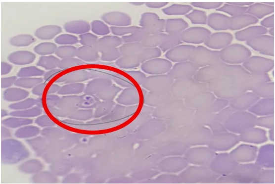
A two-year-old, intact male, Persian cat weighing 3.1 kg was presented dull and depressed to District Veterinary Centre, Palakkad, Kerala, India with history of vomiting, lethargy and progressive reduction in appetite for 4 days; the cat had a mixed indoor–outdoor lifestyle. While on examination clinically, an elevated body temperature of 103.9°F, pale conjunctival and buccal mucous membranes, dyspnea and salivation were noticed. No ticks were detected on the body of the cat during clinical examination. Haematological evaluation was performed using an automated haematology analyser (Exigo H400, Boule Diagnostics, Sweden) according to the manufacturer's instructions. The analysis revealed leukopenia, characterized by decreased neutrophil and eosinophil counts, along with an increased monocyte count. The lymphocyte count remained in the normal range. A noticeable reduction was found in total erythrocyte counts, haemoglobin and haematocrit. The red cell indices—mean corpuscular volume, mean corpuscular haemoglobin, and mean corpuscular haemoglobin concentration—remained within the established normal reference ranges, indicating a normocytic, normochromic anaemia. A severe reduction in platelet count (12×109/L) was also noticed, suggestive of thrombocytopenia. The hematological parameters observed in the present case are summarized in Table 1. Thin peripheral blood smears prepared from the ear tip were air-dried, fixed in absolute methanol for 30 seconds, and stained with Giemsa working solution for 30 minutes at room temperature. The smears were gently rinsed with buffered water, air-dried, and examined under a ×100 oil-immersion objective. Single and paired piroplasms arranged at an acute angle were observed within erythrocytes, suggestive of Babesia sp. (Fig. 1).
Table 1: Hematological parameters observed in the present case
| Blood Parameters | Reference range | Results |
|---|---|---|
| Total leukocyte Count (109/L) | 5.5-19.5 | 4.1 |
| Neutrophils (109/L) | 2.8-13 | 1.5 |
| Eosinophils (109/L) | 0.1-1 | <0.1 |
| Monocytes (109/L) | 0.2-1 | 1.2 |
| Lymphocytes (109/L) | 1-7 | 1.3 |
| Total Erythrocyte Count (1012/L) | 5-11 | 3.86 |
| Haemoglobin (g/dL) | 8-15 | 6 |
| Haematocrit (%) | 25-45 | 16.8 |
| Mean Corpuscular Volume (fL) | 39-50 | 43.6 |
| Mean Corpuscular Hemoglobin (pg) | 12.5-17.5 | 15.5 |
| Mean Corpuscular Haemoglobin Concentration (g/dL) | 31-38.5 | 35.6 |
| Platelet count (109/L) | 200-500 | 12 |
Fig. 1: Babesia sp. piroplasm in RBC on Giemsa-stained peripheral blood smear (100×).
Treatment and Evaluation
The animal was initially treated with an intramuscular injection of diminazene aceturate (Berenil®, MSD) at a dose of 3.5 mg/kg. Additionally, doxycycline was administered orally at 10 mg/kg (Doxypet, Savavet) for a duration of 21 days. Supportive care, including fluid administration, multivitamin injections, and oral haematinics, was also provided as per the requirement. The animal was handled humanely throughout the diagnostic and therapeutic procedures. All clinical interventions were performed in accordance with standard veterinary practices, with appropriate measures taken to minimize pain, stress, and suffering. Care was taken to provide appropriate supportive and therapeutic management in view of the cat's dyspnea and severe anemia. The animal's appetite began to improve from the second day of treatment and showed rapid clinical improvement. After 21 days, hematological parameters returned to normal and the cat had an uneventful recovery.
3. Results and Discussion
Feline babesiosis has been reported worldwide, with documented cases from several regions including Spain, Portugal, Israel, Poland, and Thailand.[1] Reports of babesiosis in India have been documented in regions such as Tamil Nadu, Gujarat and Himachal Pradesh.[5-7] In Kerala, multiple studies have reported the occurrence of feline babesiosis. Investigations by Sabu et al. [2013], Rejimon et al. [2022] and Vincy and Tressamol [2023] provide evidence of its prevalence across different hosts.[8-10] Furthermore, the identification of a novel feline species, Babesia panickeri, by Panicker et al. [2020] underscores the endemic nature of babesiosis in Kerala and highlights its continued presence in the region.[1]
The cat in the present case had a regular access to both indoor and outdoor environments, potentially increasing its opportunity for exposure to arthropod vectors. Clinical manifestations observed in this study, including pale mucous membranes, fever, and anorexia, are consistent with those reported in previous cases of babesiosis.[5,11,12] Similarly, the haematological alterations like anaemia, thrombocytopenia, and leukopenia align with the findings of Aziz et al. [2025] and Rejimon et al. [2022].[5,9] These changes are characteristic of babesiosis and reflect the underlying pathological processes, which commonly involve reductions in red blood cell count, haemoglobin concentration, haematocrit, and platelet count.[13] The concurrent absolute monocytosis observed in this study may reflect an increased demand for erythrophagocytic activity, resulting in increased circulating monocytes. Although leukopenia accompanied by monocytosis is rarely reported in babesiosis, this hematological finding may have been associated with immunosuppression secondary to a concurrent infection, such as Mycoplasma haemofelis and may have resolved following doxycycline administration.
Although Babesia felis is the most commonly discussed Babesia sp. in cats, several reports have documented the occurrence of large Babesia species in felines, including those associated with both cats and dogs. Since the tick was not detected from cat, the transmission of the infection remains uncertain. Although primaquine phosphate is considered efficacious and the drug of choice in feline babesiosis, its temporary unavailability necessitated the use of a combination therapy consisting of diminazene aceturate (Berenil®) and doxycycline.[5,14,15] Several species of Babesia have been reported in cats including B. felis, B. canis and B. lengau.[3,16,17] In the present case, microscopic examination revealed intraerythrocytic piroplasms morphologically suggestive of a large Babesia species. However, morphological assessment alone is insufficient for definitive species-level identification of Babesia. Therefore, molecular characterization is essential to confirm the species identity and distinguish closely related Babesia spp. The absence of molecular confirmation represents an important limitation of the present study and precludes definitive species-level identification of the organism based solely on its morphological characteristics. Thus, the present study highlights the need for low-cost molecular diagnostics in modern feline veterinary practice and explores research opportunities in endemic regions such as Kerala, where a novel Babesia species has been identified in cats.
4. Conclusions
Feline babesiosis should be considered in cats presenting with anemia, thrombocytopenia, and nonspecific clinical signs. This case emphasizes the importance of careful hematological evaluation and blood smear examination for the early detection of feline babesiosis, relevance of low-cost molecular techniques for species confirmation at veterinary practice and demonstrates the potential effectiveness of combination therapy in managing the infection.
Acknowledgement
The authors are grateful to the staff of District Veterinary Centre, Palakkad, Kerala, and to pet owner for their cooperation throughout the study.
CRediT Author Contribution Statement
Farhan Eravaramkunnath: Conceptualization; Formal Analysis; Methodology. Fathima Dhina: Conceptualization; Data Curation. Arya Ramachandran Nair Sujatha Devi: Formal Analysis; Investigation; Supervision. Bindhu Lakshmanan: Supervision, Validation, Writing – Review & editing. All authors have read and approved the final version of the manuscript for publication and agree to be accountable for all aspects of the work, ensuring that questions related to the accuracy or integrity of any part of the work are appropriately investigated and resolved.
Funding Declaration
This research did not receive any specific grant from funding agencies in the public, commercial, or not-for-profit sectors.
Institutional Review Board Statement
All procedures were conducted in accordance with institutional guidelines and applicable national animal welfare regulations. As this was a retrospective case report of routine clinical care, IACUC approval was not required. The report was prepared in accordance with the CARE guidelines.
Informed Consent Statement
Informed consent was obtained from the owner for the treatment of the animal.
Consent to Publish Statement
Informed consent was obtained from the owner for the publication of the clinical findings and associated information.
Data Availability Statement
The datasets generated and/or analyzed during the current study that support the findings are available from the corresponding author upon reasonable request.
Conflict of Interest
There is no conflict of interest.
Artificial Intelligence (AI) Use Disclosure
The authors declare that artificial intelligence (AI)-assisted tools were used only for language refinement, grammar improvement, and manuscript structuring purposes during the preparation of this work. All technical content, experimental implementation, results, and interpretations were independently developed and verified by the authors.
Supporting Information
Not applicable.
References
- [1] V. P. Panicker, A. K. Sreedharan Nair, A. Narayanan, S. George, S. V. Hameed, Molecular Identification of a Novel Species, Babesia panickeri sp. nov., from a Naturally Infected Domestic Cat of India and Its Comparison with Canine Babesia Isolates, Acta Parasitologica, 2020, 65, 913-918, doi: 10.2478/s11686-020-00228-9
- [2] A. Almendros, Y. R. Choi, P. M. Bęczkowski, K. Baiker, V. R. Barrs, J. A. Beatty, Babesia gibsoni Infection in a Cat with Immune-Mediated Haemolytic Anaemia and Thrombocytopenia, Animals, 2023, 13, 2128, doi: 10.3390/ani13132128
- [3] G. Baneth, M. J. Kenny, S. Tasker, Y. Anug, V. Shkap, A. Levy, S. E. Shaw, Infection with a Proposed New Subspecies of Babesia canis, Babesia canis subsp. presentii, in Domestic Cats, Journal of Clinical Microbiology, 2004, 42, 99-105, doi: 10.1128/jcm.42.1.99-105.2004
- [4] S. Remesar, J. L. Arnal, A. Gómez, A. Prieto, D. García-Dios, A. Benito, M. Rodríguez, G. Fernández, R. Panadero, C. López, P. Díez-Baños, P. Díaz, A Case Report of Fatal Feline Babesiosis Caused by Babesia canis in North Western Spain, BMC Veterinary Research, 2022, 18, 177, doi: 10.1186/s12917-022-03287-4
- [5] P. R. Aziz, K. Jamwal, A. Patial, D. Thakur, A Case Presentation of Babesiosis in A Mongrel Cat, Advances in Research, 2025, 26, 139-144, doi: 10.9734/air/2025/v26i41393
- [6] S. V. Mudaliar, G. R. Achary, V. S. Alwar, On a Species of Babesia in an Indian Wild Cat (Felis catus), Indian Veterinary Journal, 1950, 26, 392-395.
- [7] J. P. Varshney, P. S. Chaudhary, V. V. Deshmukh, Feline Babesiosis: A Case Report, Intas Polivet, 2008, 9, 288-291.
- [8] L. Sabu, R. Sreekrishnan, K. Devada, T. S. Rejitha, B. Lakshmanan, Babesia Species in a Cat—A Case Report, Journal of Veterinary Parasitology, 2013, 27, 68-69.
- [9] S. P. Rejimon, V. M. Mathai, S. B. Sarangom, A. Gopinath, C. S. Neethu, S. Sundaran, Piroplasmosis in a Domestic Cat-A Case Report, The Indian Veterinary Journal, 2022, 99, 66-67.
- [10] P. Vincy, P. V. Tresamol, Prevalence of Gastro-Intestinal and Haemoparasitic Infections among Domestic Cats of Kerala, Journal of Parasitic Diseases, 2023, 47, 562-565, doi: 10.1007/s12639-023-01599-2
- [11] C. Jackson, F. J. Dunning, Biliary Fever (Nuttalliosis) of the Cat: A Case in the Stellenbosch District, Journal of the South African Veterinary Medical Association, 1937, 8, 83-88.
- [12] J. McNeil, Piroplasmosis of the Domestic Cat, Journal of the South African Veterinary Medical Association, 1937, 8, 88-90.
- [13] O. M. Radostits, C. C. Gay, K. W. Hinchcliff, P. D. Constable, Veterinary Medicine: A Textbook of the Diseases of Cattle, Horses, Sheep, Pigs and Goats, 10th ed., Elsevier, 2006.
- [14] P. Abinaya, K. Rajkumar, A. A. Prabavathy, P. Vijayalakshmi, Primaquine Phosphate for Therapeutic Management of Feline Babesiosis-A Case Report, Intas Polivet, 2020, 21, 257-259.
- [15] A. L. Ayoob, J. Prittie, S. G. Hackner, Feline babesiosis, Journal of Veterinary Emergency and Critical Care, 2010, 20, 90-97, doi: 10.1111/j.1476-4431.2009.00493.x
- [16] A. M. Bosman, E. H. Venter, B. L. Penzhorn, Occurrence of Babesia felis and Babesia leo in Various Wild Felid Species and Domestic Cats in Southern Africa, based on Reverse Line Blot Analysis, Veterinary Parasitology, 2007, 144, 33-38, doi: 10.1016/j.vetpar.2006.09.025
- [17] A. M. Bosman, M. C. Oosthuizen, E. H. Venter, J. C. Steyl, T. A. Gous, B. L. Penzhorn, Babesia lengau Associated with Cerebral and Haemolytic Babesiosis in Two Domestic Cats, Parasites & Vectors, 2013, 6, 128, doi: 10.1186/1756-3305-6-128
Publisher Note
Publisher Note: The views, statements, and data in all publications solely belong to the authors and contributors. GR Scholastic is not responsible for any injury resulting from the ideas, methods, or products mentioned. GR Scholastic remains neutral regarding jurisdictional claims in published maps and institutional affiliations.
Open Access
This article is licensed under a Creative Commons Attribution-NonCommercial 4.0 International License, which permits the non-commercial use, sharing, adaptation, distribution and reproduction in any medium or format, as long as appropriate credit to the original author(s) and the source is given by providing a link to the Creative Commons License and changes need to be indicated if there are any. The images or other third-party material in this article are included in the article's Creative Commons License, unless indicated otherwise in a credit line to the material. If material is not included in the article's Creative Commons License and your intended use is not permitted by statutory regulation or exceeds the permitted use, you will need to obtain permission directly from the copyright holder. To view a copy of this License, visit: https://creativecommons.org/licenses/by-nc/4.0/
© The Author(s) 2026